|
Toll Free: (877) 796-6397 -- USA and Canada only -- |
Fax: +1-832-582-8590 Orders: +1-832-582-8158 |
Tech Support: +1-832-582-8158 Ext:3 Please provide your Order Number in the email. |
Biological Description
| Specificity | Anti-NCX1 Mouse Antibody [C23J23] detects endogenous levels of total NCX1 protein. |
|---|---|
| Background | NCX1 (sodium–calcium exchanger 1) is a ubiquitously expressed integral membrane protein crucial for cellular Ca²⁺ homeostasis, particularly in the heart and brain, where it exchanges intracellular Ca²⁺ for extracellular Na⁺ to regulate excitation–contraction coupling and neuronal signaling. Structurally, NCX1 contains nine transmembrane segments with two α-repeat regions (involved in ion binding and transport) and a large intracellular loop between TMS 5–6 housing β-repeat regions that bind regulatory Ca²⁺. It exists in multiple splice variants with tissue-specific regulatory phenotypes, with NCX1.1 being the predominant cardiac isoform. NCX1 function is modulated by local factors, ion concentrations, lipids, and phosphorylation by kinases such as PKA and PKC within a macromolecular complex containing mAKAP, phosphatases (PP1, PP2A), and other regulatory proteins. In cardiomyocytes, this organization enables precise β-adrenergic regulation, though alterations such as hyperphosphorylation and attenuated responsiveness occur in heart failure, highlighting its importance in both physiological and pathological conditions. |
Usage Information
| Application | IHC, FCM | Dilution |
|
||||
|---|---|---|---|---|---|---|---|
| Reactivity | Human | ||||||
| Source | Mouse | MW | |||||
| Storage Buffer | PBS, pH 7.2+50% Glycerol+0.05% BSA+0.01% NaN3 | Storage (from the date of receipt) |
-20°C (avoid freeze-thaw cycles), 2 years | ||||
| IHC |
Experimental Protocol:
Deparaffinization/Rehydration
1. Deparaffinize/hydrate sections:
2. Incubate sections in three washes of xylene for 5 min each.
3. Incubate sections in two washes of 100% ethanol for 10 min each.
4. Incubate sections in two washes of 95% ethanol for 10 min each.
5. Wash sections two times in dH2O for 5 min each.
6.Antigen retrieval: For Citrate: Heat slides in a microwave submersed in 1X citrate unmasking solution until boiling is initiated; continue with 10 min at a sub-boiling temperature (95°-98°C). Cool slides on bench top for 30 min.
Staining
1. Wash sections in dH2O three times for 5 min each.
2. Incubate sections in 3% hydrogen peroxide for 10 min.
3. Wash sections in dH2O two times for 5 min each.
4. Wash sections in wash buffer for 5 min.
5. Block each section with 100–400 µl of blocking solution for 1 hr at room temperature.
6. Remove blocking solution and add 100–400 µl primary antibody diluent in to each section. Incubate overnight at 4°C.
7. Remove antibody solution and wash sections with wash buffer three times for 5 min each.
8. Cover section with 1–3 drops HRPas needed. Incubate in a humidified chamber for 30 min at room temperature.
9. Wash sections three times with wash buffer for 5 min each.
10. Add DAB Chromogen Concentrate to DAB Diluent and mix well before use.
11. Apply 100–400 µl DAB to each section and monitor closely. 1–10 min generally provides an acceptable staining intensity.
12. Immerse slides in dH2O.
13. If desired, counterstain sections with hematoxylin.
14. Wash sections in dH2O two times for 5 min each.
15. Dehydrate sections: Incubate sections in 95% ethanol two times for 10 sec each; Repeat in 100% ethanol, incubating sections two times for 10 sec each; Repeat in xylene, incubating sections two times for 10 sec each.
16. Mount sections with coverslips and mounting medium.
|
References
|
Application Data
IHC
Validated by Selleck
-
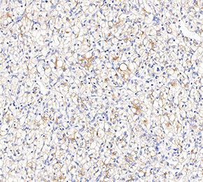 Immunohistochemical analysis of formalin fixed paraffin embedded human gastric cancer tissue with F2381 at 1:1000 dilution.
IHC
Validated by Selleck
-
Immunohistochemical analysis of formalin fixed paraffin embedded human gastric cancer tissue with F2381 at 1:1000 dilution.