|
Toll Free: (877) 796-6397 -- USA and Canada only -- |
Fax: +1-832-582-8590 Orders: +1-832-582-8158 |
Tech Support: +1-832-582-8158 Ext:3 Please provide your Order Number in the email. |
Biological Description
| Specificity | Anti-Nav1.7 Mouse Antibody [P22E18] detects endogenous levels of total Nav1.7 protein. |
|---|---|
| Background | NaV1.7 is a voltage-gated sodium channel encoded by the SCN9A gene and is predominantly expressed in peripheral sensory neurons, including small-diameter nociceptive dorsal root ganglion (DRG) neurons, trigeminal ganglion neurons, and sympathetic neurons. Structurally, NaV1.7 consists of a large α-subunit forming the ion-conducting pore, organized into four homologous domains (DI–DIV), each containing six transmembrane segments (S1–S6), with voltage-sensing regions (S4) and a selectivity filter that confers sodium specificity; it may also associate with β-subunits that modulate gating and expression. Functionally, NaV1.7 plays a crucial role in amplifying small, slow depolarizations near resting membrane potential and setting the threshold for action potential initiation, acting as a “threshold channel” in pain-sensing neurons. Gain-of-function mutations in SCN9A cause hyperexcitability of nociceptors, leading to inherited pain syndromes such as inherited erythromelalgia (IEM) and paroxysmal extreme pain disorder (PEPD), whereas loss-of-function mutations result in congenital insensitivity to pain. Through its low-threshold activation and slow inactivation kinetics, NaV1.7 tightly regulates neuronal excitability and is a major pharmacological target for pain therapeutics. |
Usage Information
| Application | IHC | Dilution |
|
||
|---|---|---|---|---|---|
| Reactivity | Mouse | ||||
| Source | Mouse | MW | |||
| Storage Buffer | PBS, pH 7.2+50% Glycerol+0.05% BSA+0.01% NaN3 | Storage (from the date of receipt) |
-20°C (avoid freeze-thaw cycles), 2 years | ||
| IHC |
Experimental Protocol:
Deparaffinization/Rehydration
1. Deparaffinize/hydrate sections:
2. Incubate sections in three washes of xylene for 5 min each.
3. Incubate sections in two washes of 100% ethanol for 10 min each.
4. Incubate sections in two washes of 95% ethanol for 10 min each.
5. Wash sections two times in dH2O for 5 min each.
6.Antigen retrieval: For Citrate: Heat slides in a microwave submersed in 1X citrate unmasking solution until boiling is initiated; continue with 10 min at a sub-boiling temperature (95°-98°C). Cool slides on bench top for 30 min.
Staining
1. Wash sections in dH2O three times for 5 min each.
2. Incubate sections in 3% hydrogen peroxide for 10 min.
3. Wash sections in dH2O two times for 5 min each.
4. Wash sections in wash buffer for 5 min.
5. Block each section with 100–400 µl of blocking solution for 1 hr at room temperature.
6. Remove blocking solution and add 100–400 µl primary antibody diluent in to each section. Incubate overnight at 4°C.
7. Remove antibody solution and wash sections with wash buffer three times for 5 min each.
8. Cover section with 1–3 drops HRPas needed. Incubate in a humidified chamber for 30 min at room temperature.
9. Wash sections three times with wash buffer for 5 min each.
10. Add DAB Chromogen Concentrate to DAB Diluent and mix well before use.
11. Apply 100–400 µl DAB to each section and monitor closely. 1–10 min generally provides an acceptable staining intensity.
12. Immerse slides in dH2O.
13. If desired, counterstain sections with hematoxylin.
14. Wash sections in dH2O two times for 5 min each.
15. Dehydrate sections: Incubate sections in 95% ethanol two times for 10 sec each; Repeat in 100% ethanol, incubating sections two times for 10 sec each; Repeat in xylene, incubating sections two times for 10 sec each.
16. Mount sections with coverslips and mounting medium.
|
References
|
Application Data
IHC
Validated by Selleck
-
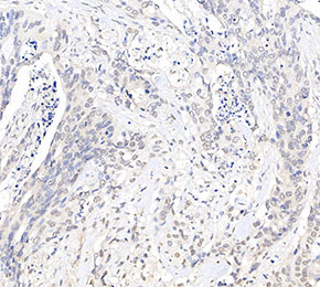 Immunohistochemical analysis of formalin fixed paraffin embedded human colorectal cancer tissue with F3300 at 1:200 dilution.