|
Toll Free: (877) 796-6397 -- USA and Canada only -- |
Fax: +1-832-582-8590 Orders: +1-832-582-8158 |
Tech Support: +1-832-582-8158 Ext:3 Please provide your Order Number in the email. |
Biological Description
| Specificity | Anti-GLP-1 Mouse Antibody [B23D23] recognizes endogenous levels of total GLP-1 protein. |
|---|---|
| Background | Glucagon-like peptide-1 (GLP-1) is a 30-amino-acid incretin hormone primarily secreted by intestinal L-cells in response to nutrient ingestion, produced through the enzymatic cleavage of the proglucagon precursor. GLP-1 contains a conserved N-terminal sequence critical for receptor activation, including key residues such as His7 that are essential for binding and signaling, and adopts α-helical conformations stabilizing its interaction with the GLP-1 receptor (GLP-1R), a G-protein-coupled receptor expressed in pancreatic β-cells, brain, gastrointestinal tract, and other tissues. Upon binding GLP-1R, the receptor activates G-proteins that stimulate adenylate cyclase, increasing intracellular cAMP, which triggers downstream effectors like protein kinase A (PKA) and Epac2, enhancing glucose-dependent insulin secretion, inhibiting glucagon release mainly via somatostatin secretion from δ-cells, and slowing gastric emptying to control postprandial glucose levels. GLP-1 also reduces appetite and promotes satiety via central nervous system pathways. GLP-1R activation exerts neuroprotective effects by modulating inflammation and promoting neuronal survival through cAMP/PKA and ERK signaling pathways. Additionally, GLP-1 influences mitochondrial function and energy metabolism in muscle and neuronal cells and favors bone formation by modulating the osteoprotegerin (OPG)/RANKL ratio to support skeletal health. Endogenously, GLP-1 is rapidly degraded by dipeptidyl peptidase-4 (DPP-4), limiting its half-life to about 1–2 minutes, prompting the development of GLP-1 receptor agonists and DPP-4 inhibitors as therapeutics for type 2 diabetes and obesity. |
Usage Information
| Application | IHC, sELISA | Dilution |
|
||
|---|---|---|---|---|---|
| Reactivity | Mouse, Human | ||||
| Source | Mouse | MW | |||
| Storage Buffer | PBS, pH 7.2+50% Glycerol+0.05% BSA+0.01% NaN3 | Storage (from the date of receipt) |
-20°C (avoid freeze-thaw cycles), 2 years | ||
| IHC |
Experimental Protocol:
Deparaffinization/Rehydration
1. Deparaffinize/hydrate sections:
2. Incubate sections in three washes of xylene for 5 min each.
3. Incubate sections in two washes of 100% ethanol for 10 min each.
4. Incubate sections in two washes of 95% ethanol for 10 min each.
5. Wash sections two times in dH2O for 5 min each.
6.Antigen retrieval: For Citrate: Heat slides in a microwave submersed in 1X citrate unmasking solution until boiling is initiated; continue with 10 min at a sub-boiling temperature (95°-98°C). Cool slides on bench top for 30 min.
Staining
1. Wash sections in dH2O three times for 5 min each.
2. Incubate sections in 3% hydrogen peroxide for 10 min.
3. Wash sections in dH2O two times for 5 min each.
4. Wash sections in wash buffer for 5 min.
5. Block each section with 100–400 µl of blocking solution for 1 hr at room temperature.
6. Remove blocking solution and add 100–400 µl primary antibody diluent in to each section. Incubate overnight at 4°C.
7. Remove antibody solution and wash sections with wash buffer three times for 5 min each.
8. Cover section with 1–3 drops HRPas needed. Incubate in a humidified chamber for 30 min at room temperature.
9. Wash sections three times with wash buffer for 5 min each.
10. Add DAB Chromogen Concentrate to DAB Diluent and mix well before use.
11. Apply 100–400 µl DAB to each section and monitor closely. 1–10 min generally provides an acceptable staining intensity.
12. Immerse slides in dH2O.
13. If desired, counterstain sections with hematoxylin.
14. Wash sections in dH2O two times for 5 min each.
15. Dehydrate sections: Incubate sections in 95% ethanol two times for 10 sec each; Repeat in 100% ethanol, incubating sections two times for 10 sec each; Repeat in xylene, incubating sections two times for 10 sec each.
16. Mount sections with coverslips and mounting medium.
|
References
|
Application Data
IHC
Validated by Selleck
-
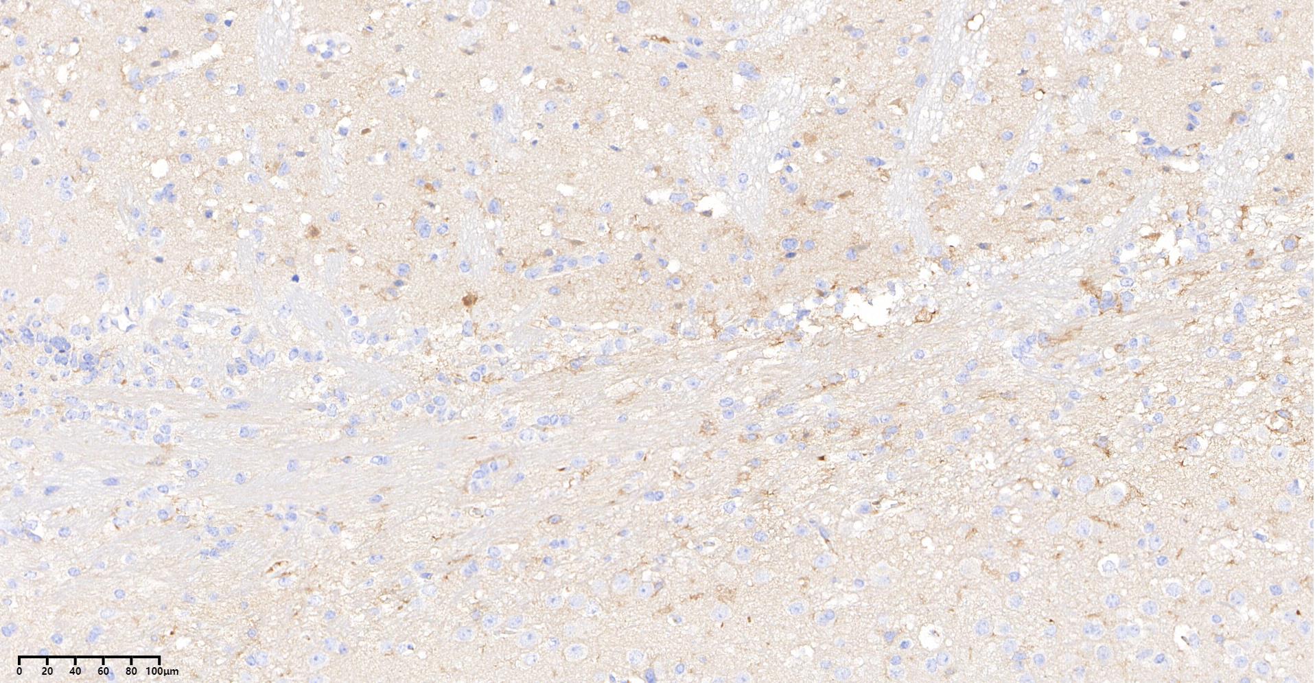 Immunohistochemical analysis of formalin fixed paraffin embedded human pancreas tissue with F1621 at 1:500 dilution.
IHC
Validated by Selleck
-
Immunohistochemical analysis of formalin fixed paraffin embedded human pancreas tissue with F1621 at 1:500 dilution.