|
Toll Free: (877) 796-6397 -- USA and Canada only -- |
Fax: +1-832-582-8590 Orders: +1-832-582-8158 |
Tech Support: +1-832-582-8158 Ext:3 Please provide your Order Number in the email. |
Biological Description
| Specificity | Anti-Endothelial Cell Mouse Antibody [K23P8] recognizes endogenous levels of total Endothelial Cell protein. |
|---|---|
| Background | Endothelial cells form a single, thin monolayer lining the interior surfaces of blood and lymphatic vessels, serving as a critical barrier between circulating fluids and surrounding tissues. They are connected by tight junctions and anchored to a basement membrane, with specialized surface proteins such as VE-cadherin and PECAM-1 that maintain vessel integrity and mediate immune cell interactions. They regulate vascular tone by releasing vasodilators like nitric oxide and vasoconstrictors such as endothelin, maintain hemostasis by balancing pro- and anticoagulant factors, and control filtration of fluids across vessel walls. They play essential roles in angiogenesis by proliferating and migrating to form new capillaries, which are important for wound healing, reproduction, and tumor growth. Endothelial cells also modulate immune surveillance through the expression of selectins and integrins, facilitating leukocyte rolling, adhesion, and transmigration during inflammation. Dysfunction of endothelial cells contributes to diseases including atherosclerosis, hypertension, diabetes-related vascular complications, and chronic inflammation. In tumors, abnormal endothelial cells form irregular, fragile vessels lacking smooth muscle and pericyte coverage, which promotes cancer progression and metastasis. Endothelial dysfunction shifts their phenotype from anticoagulant and anti-inflammatory to pro-thrombotic and pro-inflammatory, driving vascular disease progression. |
Usage Information
| Application | IHC | Dilution |
|
||
|---|---|---|---|---|---|
| Reactivity | Rat | ||||
| Source | Mouse | MW | |||
| Storage Buffer | PBS, pH 7.2+50% Glycerol+0.05% BSA+0.01% NaN3 | Storage (from the date of receipt) |
-20°C (avoid freeze-thaw cycles), 2 years | ||
| IHC |
Experimental Protocol:
Deparaffinization/Rehydration
1. Deparaffinize/hydrate sections:
2. Incubate sections in three washes of xylene for 5 min each.
3. Incubate sections in two washes of 100% ethanol for 10 min each.
4. Incubate sections in two washes of 95% ethanol for 10 min each.
5. Wash sections two times in dH2O for 5 min each.
6.Antigen retrieval: For Citrate: Heat slides in a microwave submersed in 1X citrate unmasking solution until boiling is initiated; continue with 10 min at a sub-boiling temperature (95°-98°C). Cool slides on bench top for 30 min.
Staining
1. Wash sections in dH2O three times for 5 min each.
2. Incubate sections in 3% hydrogen peroxide for 10 min.
3. Wash sections in dH2O two times for 5 min each.
4. Wash sections in wash buffer for 5 min.
5. Block each section with 100–400 µl of blocking solution for 1 hr at room temperature.
6. Remove blocking solution and add 100–400 µl primary antibody diluent in to each section. Incubate overnight at 4°C.
7. Remove antibody solution and wash sections with wash buffer three times for 5 min each.
8. Cover section with 1–3 drops HRPas needed. Incubate in a humidified chamber for 30 min at room temperature.
9. Wash sections three times with wash buffer for 5 min each.
10. Add DAB Chromogen Concentrate to DAB Diluent and mix well before use.
11. Apply 100–400 µl DAB to each section and monitor closely. 1–10 min generally provides an acceptable staining intensity.
12. Immerse slides in dH2O.
13. If desired, counterstain sections with hematoxylin.
14. Wash sections in dH2O two times for 5 min each.
15. Dehydrate sections: Incubate sections in 95% ethanol two times for 10 sec each; Repeat in 100% ethanol, incubating sections two times for 10 sec each; Repeat in xylene, incubating sections two times for 10 sec each.
16. Mount sections with coverslips and mounting medium.
|
References
|
Application Data
IHC
Validated by Selleck
-
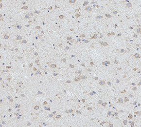 Immunohistochemical analysis of formalin fixed paraffin embedded rat brain tissue with F1713 at 1:200 dilution.