|
Toll Free: (877) 796-6397 -- USA and Canada only -- |
Fax: +1-832-582-8590 Orders: +1-832-582-8158 |
Tech Support: +1-832-582-8158 Ext:3 Please provide your Order Number in the email. |
Biological Description
| Specificity | Anti-CA19-9 Mouse Antibody [C17A13] detects endogenous levels of CA 19-9 protein. |
|---|---|
| Background | CA19-9 (Carbohydrate antigen 19-9), also known as Sialyl Lewis A (sLeᵃ), is a sialylated Lewis blood group antigen, a tetrasaccharide glycan epitope composed of sialic acid, galactose, N-acetylglucosamine, and fucose, biosynthesized via the Lewis blood group pathway and requiring a functional Lewis antigen for its formation. CA19-9 is expressed on glycoproteins and glycolipids of epithelial cell membranes, particularly in the pancreas, biliary tract, stomach, and colon, and can be shed into the bloodstream. It is the current gold-standard serum biomarker for pancreatic adenocarcinoma. Functionally, CA19-9 serves as a biomarker (for diagnosis, prognosis, monitoring treatment response, and detecting recurrence), a predictor (correlating with tumor burden, stage, and resectability), and a promoter of cancer progression by facilitating E-selectin–mediated adhesion, enhancing angiogenesis, modulating immune responses, and influencing tumor microenvironment interactions. Beyond pancreatic cancer, CA19-9 is elevated in other gastrointestinal malignancies and certain benign diseases, and is under investigation as a therapeutic target via antibodies, vaccines, biosynthesis inhibitors, and CA19-9–guided drug delivery systems. |
Usage Information
| Application | IHC, FCM, ELISA | Dilution |
|
||
|---|---|---|---|---|---|
| Reactivity | Human | ||||
| Source | Mouse | MW | |||
| Storage Buffer | PBS, pH 7.2+50% Glycerol+0.05% BSA+0.01% NaN3 | Storage (from the date of receipt) |
-20°C (avoid freeze-thaw cycles), 2 years | ||
| IHC |
Experimental Protocol:
Deparaffinization/Rehydration
1. Deparaffinize/hydrate sections:
2. Incubate sections in three washes of xylene for 5 min each.
3. Incubate sections in two washes of 100% ethanol for 10 min each.
4. Incubate sections in two washes of 95% ethanol for 10 min each.
5. Wash sections two times in dH2O for 5 min each.
6.Antigen retrieval: For Citrate: Heat slides in a microwave submersed in 1X citrate unmasking solution until boiling is initiated; continue with 10 min at a sub-boiling temperature (95°-98°C). Cool slides on bench top for 30 min.
Staining
1. Wash sections in dH2O three times for 5 min each.
2. Incubate sections in 3% hydrogen peroxide for 10 min.
3. Wash sections in dH2O two times for 5 min each.
4. Wash sections in wash buffer for 5 min.
5. Block each section with 100–400 µl of blocking solution for 1 hr at room temperature.
6. Remove blocking solution and add 100–400 µl primary antibody diluent in to each section. Incubate overnight at 4°C.
7. Remove antibody solution and wash sections with wash buffer three times for 5 min each.
8. Cover section with 1–3 drops HRPas needed. Incubate in a humidified chamber for 30 min at room temperature.
9. Wash sections three times with wash buffer for 5 min each.
10. Add DAB Chromogen Concentrate to DAB Diluent and mix well before use.
11. Apply 100–400 µl DAB to each section and monitor closely. 1–10 min generally provides an acceptable staining intensity.
12. Immerse slides in dH2O.
13. If desired, counterstain sections with hematoxylin.
14. Wash sections in dH2O two times for 5 min each.
15. Dehydrate sections: Incubate sections in 95% ethanol two times for 10 sec each; Repeat in 100% ethanol, incubating sections two times for 10 sec each; Repeat in xylene, incubating sections two times for 10 sec each.
16. Mount sections with coverslips and mounting medium.
|
References
|
Application Data
IHC
Validated by Selleck
-
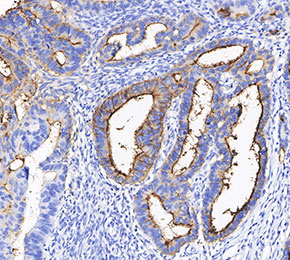 Immunohistochemical analysis of formalin fixed paraffin embedded human colorectal cancer tissue with F2466 at 1:100 dilution.