|
Toll Free: (877) 796-6397 -- USA and Canada only -- |
Fax: +1-832-582-8590 Orders: +1-832-582-8158 |
Tech Support: +1-832-582-8158 Ext:3 Please provide your Order Number in the email. |
Biological Description
| Specificity | Anti-ATP-binding cassette sub-family A member 3 Mouse Antibody [F19C21] detects endogenous levels of total ATP-binding cassette sub-family A member 3 protein. |
|---|---|
| Background | ATP-binding cassette sub-family A member 3 (ABCA3) is a highly conserved, multi-pass transmembrane glycoprotein in the ABC transporter superfamily that uses ATP hydrolysis to transport phospholipids into lamellar bodies (LBs) of alveolar type II cells, where pulmonary surfactant is assembled and stored. Structurally, ABCA3 consists of two similar halves, each containing six transmembrane domains and a cytosolic nucleotide-binding domain (NBD) with Walker A, Walker B, and ABCA-specific signature motifs, plus two essential N-linked glycosylation sites at Asn124 and Asn140 required for proper folding, stability, and LB targeting. Although expressed in multiple tissues, ABCA3 is highly enriched in the lung, where it is critical for LB biogenesis and surfactant homeostasis, particularly in transporting phosphatidylcholine, phosphatidylglycerol, and cholesterol. Mutations in ABCA3 can cause trafficking or functional defects, leading to neonatal surfactant deficiency, interstitial lung disease, and fatal respiratory distress. |
Usage Information
| Application | IHC | Dilution |
|
||
|---|---|---|---|---|---|
| Reactivity | Mouse, Rat | ||||
| Source | Mouse | MW | |||
| Storage Buffer | PBS, pH 7.2+50% Glycerol+0.05% BSA+0.01% NaN3 | Storage (from the date of receipt) |
-20°C (avoid freeze-thaw cycles), 2 years | ||
| IHC |
Experimental Protocol:
Deparaffinization/Rehydration
1. Deparaffinize/hydrate sections:
2. Incubate sections in three washes of xylene for 5 min each.
3. Incubate sections in two washes of 100% ethanol for 10 min each.
4. Incubate sections in two washes of 95% ethanol for 10 min each.
5. Wash sections two times in dH2O for 5 min each.
6.Antigen retrieval: For Citrate: Heat slides in a microwave submersed in 1X citrate unmasking solution until boiling is initiated; continue with 10 min at a sub-boiling temperature (95°-98°C). Cool slides on bench top for 30 min.
Staining
1. Wash sections in dH2O three times for 5 min each.
2. Incubate sections in 3% hydrogen peroxide for 10 min.
3. Wash sections in dH2O two times for 5 min each.
4. Wash sections in wash buffer for 5 min.
5. Block each section with 100–400 µl of blocking solution for 1 hr at room temperature.
6. Remove blocking solution and add 100–400 µl primary antibody diluent in to each section. Incubate overnight at 4°C.
7. Remove antibody solution and wash sections with wash buffer three times for 5 min each.
8. Cover section with 1–3 drops HRPas needed. Incubate in a humidified chamber for 30 min at room temperature.
9. Wash sections three times with wash buffer for 5 min each.
10. Add DAB Chromogen Concentrate to DAB Diluent and mix well before use.
11. Apply 100–400 µl DAB to each section and monitor closely. 1–10 min generally provides an acceptable staining intensity.
12. Immerse slides in dH2O.
13. If desired, counterstain sections with hematoxylin.
14. Wash sections in dH2O two times for 5 min each.
15. Dehydrate sections: Incubate sections in 95% ethanol two times for 10 sec each; Repeat in 100% ethanol, incubating sections two times for 10 sec each; Repeat in xylene, incubating sections two times for 10 sec each.
16. Mount sections with coverslips and mounting medium.
|
References
|
Application Data
IHC
Validated by Selleck
-
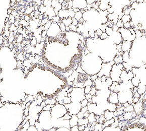 Immunohistochemical analysis of formalin fixed paraffin embedded mouse lung tissue with F2545 at 1:100 dilution.