OSI-930

Catalog No.S1220 Batch:S122001

Print

Technical Data

Formula

C22H16F3N3O2S

Molecular Weight 443.44 CAS No. 728033-96-3
Solubility (25°C)* In vitro DMSO 89 mg/mL (200.7 mM)
Ethanol 3 mg/mL (6.76 mM)
Water Insoluble
In vivo (Add solvents to the product individually and in order)
Homogeneous suspension
CMC-NA
≥5mg/ml Taking the 1 mL working solution as an example, add 5 mg of this product to 1 ml of CMC-Na solution, mix evenly to obtain a homogeneous suspension with a final concentration of 5 mg/ml.
* <1 mg/ml means slightly soluble or insoluble.
* Please note that Selleck tests the solubility of all compounds in-house, and the actual solubility may differ slightly from published values. This is normal and is due to slight batch-to-batch variations.
* Room temperature shipping (Stability testing shows this product can be shipped without any cooling measures.)

Preparing Stock Solutions

Biological Activity

Description OSI-930 is a potent inhibitor of Kit (c-Kit), KDR and CSF-1R with IC50 of 80 nM, 9 nM and 15 nM, respectively; also potent to Flt-1, c-Raf and Lck and low activity against PDGFRα/β, Flt-3 and Abl. Phase 1.
Targets
FLT1 [1]
(Cell-free assay)
KDR [1]
(Cell-free assay)
CSF-1R [1]
(Cell-free assay)
LCK [1]
(Cell-free assay)
C-Raf [1]
(Cell-free assay)
View More
8 nM 9 nM 15 nM 22 nM 41 nM
In vitro OSI-930 inhibits the cell proliferation in the HMC-1 cell line with IC50 of 14 nM without significant effect on growth of the COLO-205 cell line that does not express a constitutively active mutant receptor tyrosine kinase. Moreover, OSI-930 also induces apoptosis in HMC-1 cell line with EC50 of 34 nM. [1] A recent study shows that OSI-930 inactivates purified, recombinant cytochrome P450 (P450) 3A4 with a Ki of 24 μM in a time- and concentration-dependent mode. [2]
In vivo OSI-930, administrated at the maximally efficacious dose of 200 mg/kg by oral gavage, exhibits potent antitumor activity in a broad range of preclinical xenograft models including HMC-1, NCI-SNU-5, COLO-205 and U251 xenograft models. [1]

Protocol (from reference)

Kinase Assay:[1]
  • Protein kinase assays

    Protein kinase assays are either done in-house by ELISA-based assay methods (Kit, KDR, PDGFRα, and PDGFRβ) or by a radiometric method. In-house ELISA assays used poly(Glu:Tyr) as the substrate bound to the surface of 96-well assay plates; phosphorylation is then detected using an antiphosphotyrosine antibody conjugated to HRP. The bound antibody is then quantitated using ABTS as the peroxidase substrate by measuring the absorbance at 405/490 nm. All assays uses purified recombinant kinase catalytic domains that are either expressed in insect cells or in bacteria. The Kit and EGFR protein used for in-house assays are prepared internally; other enzymes are obtained. Recombinant Kit protein is expressed as an NH2-terminal glutathione S-transferase fusion protein in insect cells and is initially purified as a nonphosphorylated (nonactivated) enzyme with a relatively high Km for ATP (400 μM). In some assays, an activated (tyrosine phosphorylated) form of the enzyme is prepared by incubation with 1 mM ATP for 1 hour at 30 °C. The phosphorylated protein is then passed through a desalting column to remove the majority of the ATP and stored at −80 °C in buffer containing 50% glycerol. The resultant preparation has a considerably higher specific activity and a lower Km for ATP (25 μM) than the initial nonphosphorylated preparation. The inhibition of Kit autophosphorylation by OSI-930 is assayed by incubation of the nonphosphorylated enzyme at 30 °C in the presence of 200 μM ATP and various concentrations of OSI-930. The reaction is stopped by removal of aliquots into SDS-PAGE sample buffer followed by heating to 100 °C for 5 minutes. The degree of phosphorylation of Kit is then determined by immunoblotting for both total Kit and phosphorylated Kit.

Cell Assay:[1]
  • Cell lines

    HMC-1 and COLO-205

  • Concentrations

    0--1 μM

  • Incubation Time

    48 hours

  • Method

    For assays of cell proliferation and apoptosis, cells are seeded into 96-well plates and incubated for 2 to 3 days in the presence of OSI-930 at various concentrations. Inhibition of cell growth is determined by luminescent quantitation of the intracellular ATP content using CellTiterGlo. Induction of caspase-dependent apoptosis by OSI-930 is quantitated by an enzymatic caspase 3/7 assay. Inhibition of angiogenesis by OSI-930 is monitored using the rat aortic ring endothelial sprout outgrowth assay. Sections of aorta are prepared from CO2-euthanized male rats and cultured in vitro in a collagen matrix in the presence or absence of OSI-930. The collagen matrix is prepared from type 1 rat tail collagen solubilized in 0.1% acetic acid at 3 mg/mL, which is combined with 0.125 volume collagen buffer (0.05 N NaOH, 200 mM HEPES, 260 mM NaHCO3), 0.125 volume of medium 199, 0.0125 volume of 1 M NaOH, and 1% GlutaMax. Aortic rings are embedded in 0.4 mL of this matrix in six-well plates, to which 0.5 mL endothelial basal medium and the appropriate amount of OSI-930 is added; the rings are then incubated for 10 days and the resultant angiogenic sprout outgrowth is digitally quantitated from images by measurement of the sprout-containing area within a series of concentric rings around the aortic tissue area.

Animal Study:[1]
  • Animal Models

    HMC-1, NCI-SNU-5, COLO-205 and U251 cells are injected s.c. into the right flank of CD1 nu/nu mice.

  • Dosages

    ≤200 mg/kg

  • Administration

    Administered via p.o.

References

  • https://pubmed.ncbi.nlm.nih.gov/16424037/
  • https://pubmed.ncbi.nlm.nih.gov/21068193/

Customer Product Validation

RE-luc2P-HEK293 cells were pretreated with 1uM OSI-930 (green), 20uM TBB (blue), 10uM CKI-7 (purple), or 10uM H-89 (orange) for 16 h and infected with Y. enterocolitica WA or Y. pestis Ind195 at MOI 1 and 20, respectively, for 1 h. Following stimulation with 10 ng/ml TNF-α at 5 h post-infection, luciferase activity was measured 24 h post-infection. Results were determined from two independent experiments performed in triplicate. A"*" denotes that the % NF-κβ inhibition using the inhibitors was significantly different (p<0.05) compared to the no drug control (black). The relative NF-κB inhibition by Yersinia infection was determined as a percentage of luciferase activity in bacteria-infected cells relative to luciferase activity in bacteria-free control cells.

Data from [ BMC Microbiol , 2013 , 13, 249 ]

<p> </p><p>Table 2. shows the reversal effect of OSI-930 and Fumitremorgin C (FTC) on the cytotoxicity of mitoxantrone to HEK293/pcDNA3.1 and HEK293-ABCG2-482-R2.<sup>a</sup>IC<sub>50</sub>: concentration that inhibited cell survival by 50% (means ± SD). <sup>b</sup>FR: fold-resistance was the value of that IC<sub>50</sub> value for mitoxantrone of HEK293/pcDNA3.1 cells was divided by IC<sub>50</sub> value for mitoxantrone of HEK293/pcDNA3.1 and HEK293-ABCG2-482-R2 cells in the absence or presence of OSI-930 and Fumitremorgin C (FTC). Values in table are representative of at least three independent experiments performed in triplicate.</p>

,

<p>For MTT assays, cells (2,000 ~ 5,000 cells/well) were subcultured into 96-well plates according to their growth properties. Cell proliferation was assayed at 72 hr after treatment of OSI-930 by adding 20 μl of 5 mg/ml 3-(4,5-Dimethylthiazol-2-yl)-2,5-diphenyltetrazolium bromide (MTT) solution per 100 μl of growth medium. After incubating for 3-4 h at 37°C, the media were removed and 150 µl/well of MTT solvent (either absolute DMSO or isopropanol containing 4 mM HCl and 0.1% Nonidet-40) was added to dissolve the formazan</p>

, , Dr. Yong-Weon Yi from Georgetown University Medical Center

Selleck's OSI-930 has been cited by 8 publications

Orthogonal proteogenomic analysis identifies the druggable PA2G4-MYC axis in 3q26 AML [ Nat Commun, 2024, 15(1):4739] PubMed: 38834613
Small-Molecule and CRISPR Screening Converge to Reveal Receptor Tyrosine Kinase Dependencies in Pediatric Rhabdoid Tumors. [ Cell Rep, 2019, 28(9):2331-2344] PubMed: 31461650
TLR7/8-agonist-loaded Nanoparticles Promote the Polarization of Tumour-Associated Macrophages to Enhance Cancer Immunotherapy [ Nat Biomed Eng, 2018, 2(8):578-588] PubMed: 31015631
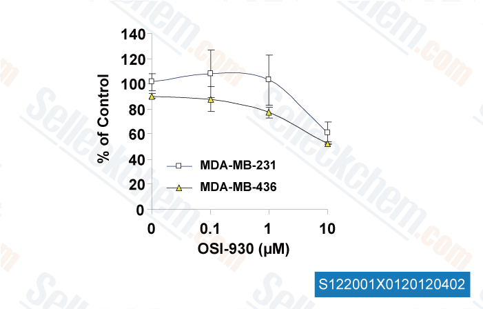
Targeting a cell state common to triple-negative breast cancers [Muellner MK, et al. Mol Syst Biol, 2015, 11(1):789] PubMed: 25699542
Dual inhibition of EGFR and MET induces synthetic lethality in triple-negative breast cancer cells through downregulation of ribosomal protein S6 [ Int J Oncol, 2015, 47(1):122-32] PubMed: 25955731
Dual inhibition of EGFR and MET induces synthetic lethality in triple-negative breast cancer cells through downregulation of ribosomal protein S6. [Yi YW, et al. Int J Oncol, 2015, 47(1):122-32] PubMed: 25955731
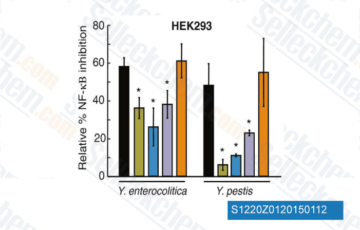
c-KIT signaling is targeted by pathogenic Yersinia to suppress the host immune response. [Micheva-Viteva SN, et al. BMC Microbiol, 2013, 13(1):249] PubMed: 24206648
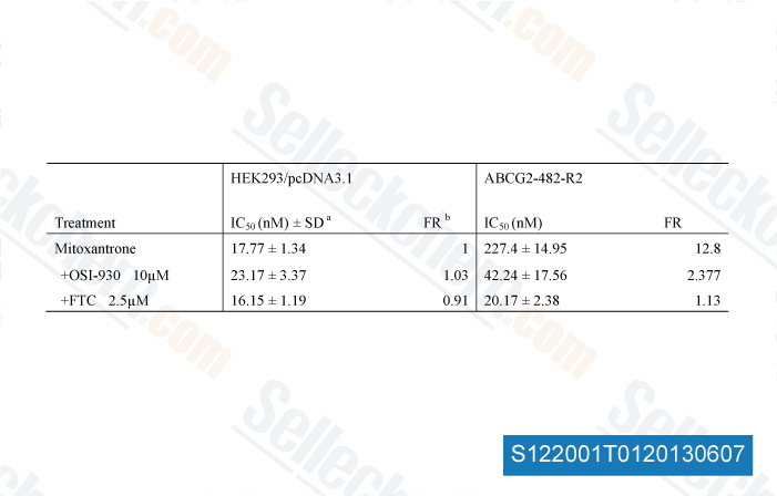
OSI-930 analogues as novel reversal agents for ABCG2-mediated multidrug resistance. [Kuang Y, et al. Biochem Pharmacol, 2012, 84(6):766-74] PubMed: 22750060

RETURN POLICY
Selleck Chemical’s Unconditional Return Policy ensures a smooth online shopping experience for our customers. If you are in any way unsatisfied with your purchase, you may return any item(s) within 7 days of receiving it. In the event of product quality issues, either protocol related or product related problems, you may return any item(s) within 365 days from the original purchase date. Please follow the instructions below when returning products.

SHIPPING AND STORAGE
Selleck products are transported at room temperature. If you receive the product at room temperature, please rest assured, the Selleck Quality Inspection Department has conducted experiments to verify that the normal temperature placement of one month will not affect the biological activity of powder products. After collecting, please store the product according to the requirements described in the datasheet. Most Selleck products are stable under the recommended conditions.

NOT FOR HUMAN, VETERINARY DIAGNOSTIC OR THERAPEUTIC USE.