MK-2206 2HCl

Catalog No.S1078 Batch:S107813

Print

Technical Data

Formula

C25H21N5O.2HCl

Molecular Weight 480.39 CAS No. 1032350-13-2
Solubility (25°C)* In vitro DMSO 96 mg/mL (199.83 mM)
Water 10 mg/mL (20.81 mM)
Ethanol Insoluble
In vivo (Add solvents to the product individually and in order)
Homogeneous suspension
CMC-NA
≥5mg/ml Taking the 1 mL working solution as an example, add 5 mg of this product to 1 ml of CMC-Na solution, mix evenly to obtain a homogeneous suspension with a final concentration of 5 mg/ml.
Clear solution
5%DMSO 40%PEG300 5%Tween80 50%ddH2O

Validated by Selleck labs. Should you need adjustments to this formulation, contact our sales team for custom testing.

4.800mg/ml (9.99mM) Taking the 1 mL working solution as an example, add 50 μL of 96 mg/ml clarified DMSO stock solution to 400 μL PEG300, mix evenly to clarify it; add 50 μL Tween80 to the above system, mix evenly to clarify it; then continue to add 500 μL ddH2O to adjust the volume to 1 mL. The mixed solution should be used immediately for optimal results. 
Clear solution
5% DMSO 95% corn oil

Validated by Selleck labs. Should you need adjustments to this formulation, contact our sales team for custom testing.

0.400mg/ml (0.83mM) Taking the 1 mL working solution as an example, add 50 μL of 8 mg/ml clear DMSO stock solution to 950 μL of corn oil and mix evenly. The mixed solution should be used immediately for optimal results. 
* <1 mg/ml means slightly soluble or insoluble.
* Please note that Selleck tests the solubility of all compounds in-house, and the actual solubility may differ slightly from published values. This is normal and is due to slight batch-to-batch variations.
* Room temperature shipping (Stability testing shows this product can be shipped without any cooling measures.)

Preparing Stock Solutions

Biological Activity

Description MK-2206 2HCl is a highly selective inhibitor of Akt1/2/3 with IC50 of 8 nM/12 nM/65 nM in cell-free assays, respectively; no inhibitory activities against 250 other protein kinases observed. MK-2206 2HCl induces autophagy and apoptosis in cancer cells. Phase 2.
Targets
Akt1 [1]
(Cell-free assay)
Akt2 [1]
(Cell-free assay)
Akt3 [1]
(Cell-free assay)
8 nM 12 nM 65 nM
In vitro

MK-2206 is an allosteric inhibitor and is activated by the pleckstrin homology domain. MK-2206 inhibits auto-phosphorylation of both Akt T308 and S473. MK-2206 also prevents Akt-mediated phosphorylation of downstream signaling molecules, including TSC2, PRAS40 and ribosomal S6 proteins. [1] MK-2206 inhibits Ras wild-type (WT) cell lines (A431, HCC827, and NCI-H292) more potently when compared to Ras-mutant cell lines (NCI-H358, NCI-H23, NCI-H1299, and Calu-6). MK-2206 also shows synergistic responses in combination with cytotoxic agents in lung NCI-H460 or ovarian A2780 tumor cells. [2] MK-2206 or siRNA-mediated Akt inhibition strongly activates autophagy in human glioma cells. However, eukaryotic elongation factor-2 (eEF-2) silencing suppresses MK-2206-induced-autophagy, with a promotion of apoptotic cell death. [3]

In vivo

MK-2206 shows 60% TGI and inhibits more than 70 % of phospho-Akt1/2 (T308 and S473) in A2780 ovarian cancer xenografts at a dose of 240 mg/kg. [1] MK-2206 exhibits significant antitumor activity in NCI-H292 xenograft in combination. [2]

Features The first allosteric small molecule inhibitor of Akt to enter clinical development.

Protocol (from reference)

Kinase Assay:

[4]

  • Akt kinases assay

    Akt kinases are assayed by a GSK-derived biotinylated peptide substrate. The extent of peptide phosphorylation is determined by Homogeneous Time Resolved Fluorescence (HTRF) using a lanthanide chelate (Lance)-coupled monoclonal antibody specific for the phosphopeptide in combination with a streptavidin-linked allophycocyanin (SA-APC) fluorophore which will bind to the biotin moiety on the peptide. When the Lance and APC are in proximity, a non-radiative energy transfer takes place from the Lance to the APC, followed by emission of light from APC at 655 nm. 10X assay buffer: 500 mM HEPES, pH7.5, 1% PEG, 16.6 mM EDTA, 1 mM EGTA, 1% BSA, 20 mM 9-glycerol phosphate; Quench buffer 50 mM HEPES pH 7.3, 16.6 mM EDTA, 0.1% BSA, 0.1% Triton X-100, 0.17 nM labeled monoclonal antibody, 0.0067 mg/mL SA-APC; ATP/MgCl2 working solution: 1X Assay buffer, 1 mM DTT, 1X PIC, 5% glycerol, active Akt; Peptide working solution: 1X Assay buffer, 1 mM DTT, 1X PIC, 5% glycerol, 2 TM GSK biotinylated peptide. The reaction is assembled by adding 16 µL of ATP/MgCl2 working solution to the appropriate wells. MK-2206 or vehicle (1.0 µL) is added followed by 10 µL of peptide working solution. The reaction is started by adding 13 μL of the enzyme working solution and mixing. The reaction is allowed to proceed for 50 min and then stopped by the addition of 60 µL HTRF quench buffer. The stopped reactions are incubated at room temperature for at least 30 min and then read in the instrument.

Cell Assay:

[2]

  • Cell lines

    A431, HCC827, NCI-H292, NCI-H358, NCI-H23, NCI-H1299, Calu-6 and NCI-H460 cells

  • Concentrations

    0, 0.3, 1 and 3 μM

  • Incubation Time

    72 or 96 hours

  • Method

    MK-2206 is dissolved in DMSO as a stock solution and diluted by culture media before use. Cells are seeded at a density of 2-3 × 10³ in 96-well plates and incubated for 24 hours. Then MK-2206 (0, 0.3, 1 and 3 μM) is added to the cells. Cell proliferation is determined after 72 or 96 hours.

Animal Study:

[2]

  • Animal Models

    SK-OV-3, NCI-H292, HCC70, PC-3, and NCI-H460 models in male CD1-nude mice

  • Dosages

    120 mg/kg

  • Administration

    Orally administered

References

  • http://cancerres.aacrjournals.org/content/69/9_Supplement/DDT01-1
  • https://pubmed.ncbi.nlm.nih.gov/20571069/
  • https://pubmed.ncbi.nlm.nih.gov/21307130/
  • http://worldwide.espacenet.com/publicationDetails/biblio?CC=WO&NR=2008070016A2&KC=A2&FT=D&ND=5&date=

Customer Product Validation

VE-cadherin-induced Akt activation mediates YAP phosphorylation and translocation in ECs. HUVECs were starved for 1h and treated with thrombin (1U) for 1h. Total cell lysates were probed with anti-pAkt, Akt or b-actin antibody. The representative blots of three independent experiments are depicted, and the normalized values for p-Akt are shown. HUVECs were cultured and starved as described as in d and incubated for 8h in complete medium with the Akt inhibitor, MK-2206 (1 uM). pAkt, Akt, pYAP and YAP were detected by western blotting using specific antibodies.

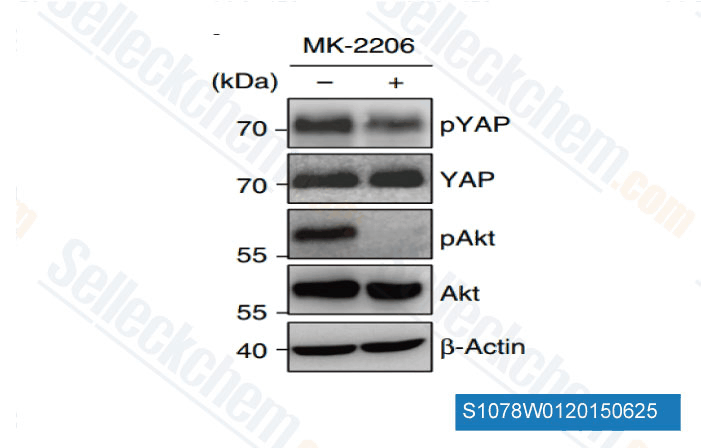
Data from [ Nat Commun , 2015 , 6:6943 ]

Inhibitors of AKT or ERK overcome SDF-1a-mediated resistance to ibrutinib-triggered PARP and caspase 3 cleavage in CXCR4S338X-expressing BCWM.1 cells. CXCR4S338X-expressing WM cells were treated with ibrutinib (0.5 uM) alone or in the presence of SDF-1a (20 nM) and/or the AKT inhibitors MK-2206 (0.5 uM) and AZD-5363 (0.5 uM); or the MEK inhibitors AS-703026 (0.25 uM), AZD-6244 (0.5 uM) and UO126 (5.0 uM). (a) Immunoblotting results for phosphoAKT (S473) and phospho-ERK (T202/Y204) in CXCR4S338X-expressing BCWM.1 cells pretreated with ibrutinib with and without AKT or ERK inhibitors, then subjected to SDF-1a stimulation for 2 min. The inhibitory effect of AZD-5363 on AKT, which is known to paradoxically hyper-phosphorylate pAKT(S473) was confirmed by inhibition of the phospho-activity for the downstream AKT targets glycogen synthase kinase 3b and pS6. (b) Immunoblotting results for cleaved PARP and cleaved caspase 3 in CXCR4S338X-expressing BCWM.1 cells treated with ibrutinib and/or AKT or ERK inhibitors for 6 h at IC50 doses. GAPDH, glyceraldehyde 3-phosphate dehydrogenase.

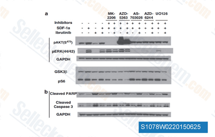
Data from [ Leukemia , 2015 , 29(1), 169-76 ]

Rap1b negatively regulates neutrophil transcellular migration by limiting PI3K-Akt signaling. (A-D) Effect of Akt inhibitor MK2206 (2 uM), Src inhibitor PP2 (10 uM), or vehicle control (DMSO) on WT or Rap1b-/- neutrophil functions. (A) Percentage of neutrophil transendothelial migration in 3D migration model. (B) ECM degradation assessed on Oregon green-labeled gelatin matrix; (left) representative images on (bar, 10 um) and (right) bar graph is percentage of matrix degradation. (C) Percentage of cells forming multiple protrusions. (D) Percentage of neutrophils present at junction of activated bEND.3 in 3D migration assay. Mean ?SD; n = 3 independent experiments. **, P < 0.01; ***, P < 0.001; NS, not significant using unpaired Student

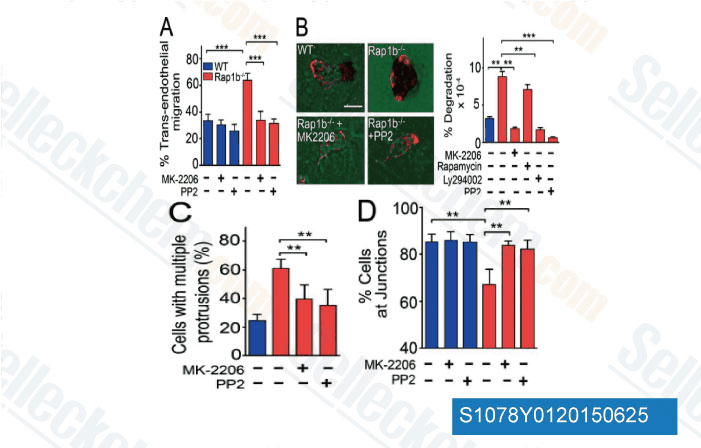
Data from [ J Exp Med , 2014 , 211(9), 1741-58 ]

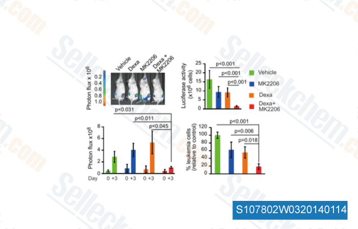
Data from [ Cancer Cell , 2013 , 24, 766-76 ]

Selleck's MK-2206 2HCl Has Been Cited by 2187 Publications

Lysosomal EGFR acts as a Rheb-GEF independent of its kinase activity to activate mTORC1 [ Cell Res, 2025, 10.1038/s41422-025-01110-x] PubMed: 40259053
Oncogenic RAS induces a distinctive form of non-canonical autophagy mediated by the P38-ULK1-PI4KB axis [ Cell Res, 2025, 10.1038/s41422-025-01085-9] PubMed: 40055523
Chromosome mis-segregation triggers cell cycle arrest through a mechanosensitive nuclear envelope checkpoint [ Nat Cell Biol, 2025, 27(1):73-86] PubMed: 39779939
Nucleus-translocated glucokinase functions as a protein kinase to phosphorylate TAZ and promote tumour growth [ Nat Commun, 2025, 16(1):7156] PubMed: 40759645
RIOK3 mediates the degradation of 40S ribosomes [ Mol Cell, 2025, 85(4):802-814.e12] PubMed: 39947183
CD24 Regulates the Formation of Ectosomes in B Lymphocytes [ J Extracell Vesicles, 2025, 14(5):e70093] PubMed: 40415253
PIP5K1A Suppresses Ferroptosis and Induces Sorafenib Resistance by Stabilizing NRF2 in Hepatocellular Carcinoma [ Adv Sci (Weinh), 2025, 12(30):e04372] PubMed: 40405713
EGFR TKIs suppress MUC1 glycosylation through the PI3K/AKT/SP1/C1GALT1 pathway to enhance TnMUC1 CAR-T efficacy in EGFR-mutant NSCLC [ Cell Rep Med, 2025, S2666-3791(25)00272-1] PubMed: 40562040
The PLEKHA1-TACC2 fusion gene drives tumorigenesis via vascular mimicry formation in esophageal squamous-cell carcinoma [ Cell Death Differ, 2025, 10.1038/s41418-025-01536-1] PubMed: 40615663
Ferroptosis in acute liver Failure: Unraveling the hepcidin-ferroportin axis and therapeutic interventions [ Redox Biol, 2025, 84:103657] PubMed: 40393152

RETURN POLICY
Selleck Chemical’s Unconditional Return Policy ensures a smooth online shopping experience for our customers. If you are in any way unsatisfied with your purchase, you may return any item(s) within 7 days of receiving it. In the event of product quality issues, either protocol related or product related problems, you may return any item(s) within 365 days from the original purchase date. Please follow the instructions below when returning products.

SHIPPING AND STORAGE
Selleck products are transported at room temperature. If you receive the product at room temperature, please rest assured, the Selleck Quality Inspection Department has conducted experiments to verify that the normal temperature placement of one month will not affect the biological activity of powder products. After collecting, please store the product according to the requirements described in the datasheet. Most Selleck products are stable under the recommended conditions.

NOT FOR HUMAN, VETERINARY DIAGNOSTIC OR THERAPEUTIC USE.