|
Toll Free: (877) 796-6397 -- USA and Canada only -- |
Fax: +1-832-582-8590 Orders: +1-832-582-8158 |
Tech Support: +1-832-582-8158 Ext:3 Please provide your Order Number in the email. |
Technical Data
| Formula | C17H14O4 |
||||||||||
| Molecular Weight | 282.29 | CAS No. | 117570-53-3 | ||||||||
| Solubility (25°C)* | In vitro | DMSO | 10.9 mg/mL (38.61 mM) | ||||||||
| Water | Insoluble | ||||||||||
| Ethanol | Insoluble | ||||||||||
| In vivo (Add solvents to the product individually and in order) |
|
||||||||||
|
* <1 mg/ml means slightly soluble or insoluble. * Please note that Selleck tests the solubility of all compounds in-house, and the actual solubility may differ slightly from published values. This is normal and is due to slight batch-to-batch variations. * Room temperature shipping (Stability testing shows this product can be shipped without any cooling measures.) |
|||||||||||
Preparing Stock Solutions
Biological Activity
| Description | Vadimezan (DMXAA) is a vascular disrupting agents (VDA) and competitive inhibitor of DT-diaphorase with Ki of 20 μM and IC50 of 62.5 μM in cell-free assays, respectively. DMXAA (Vadimezan) is also a STING agonist with potential antineoplastic activity. DMXAA (Vadimezan) potently induces IFN-β but relatively low TNF-α expression in vitro. DMXAA (Vadimezan) has antiviral activity. Phase 3. | ||||
|---|---|---|---|---|---|
| Targets |
|
||||
| In vitro | In DLD-1 human colon carcinoma cells, DMXAA inhibits DT-diaphorase activity without significant effects on the activity of cytochrome b5 reductase and cytochrome P450 reductase. Combination of menadione and DMXAA leads to an increase in the antiproliferative activity of DLD-1 cells. [1] DMXAA, as an antiviral agent, inhibits VSV-induced cytotoxicity and influenza virus replication in RAW 264.7 macrophages. [2] A recent study shows that DMXAA has non-immune-mediated inhibitory effects against several kinase members of VEGFR (vascular endothelial growth factor receptor), such as VEGFR2 signalling in human umbilical vein endothelial cells. [3] | ||||
| In vivo | DMXAA treatment significantly protects C57BL/6J mice infected i.n. with 200 p.f.u. mouse-adapted H1N1 influenza PR8 virus with 60% survival, while the control group only exhibited 20% survival. [2] DMXAA significantly delays tumor growth induced by chemical carcinogen, increases the time to tumor doubling and increases time from treatment to euthanasia. After the treatment of DMXAA, median tumor doubling time, median tumour tripling time and median time from treatment to euthanasia in tumor-bearing animals are increased by approximately 4.4-, 1.8- and 2.7-fold, respectively. [4] |
Protocol (from reference)
| Kinase Assay:[1] |
|
|---|---|
| Cell Assay:[1] |
|
| Animal Study:[4] |
|
References
|
Customer Product Validation
-
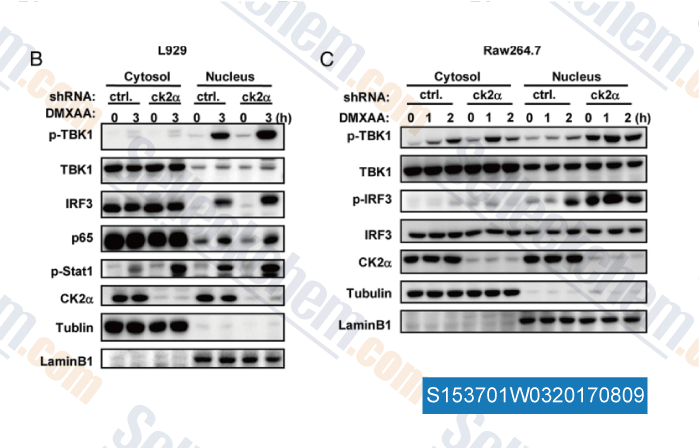
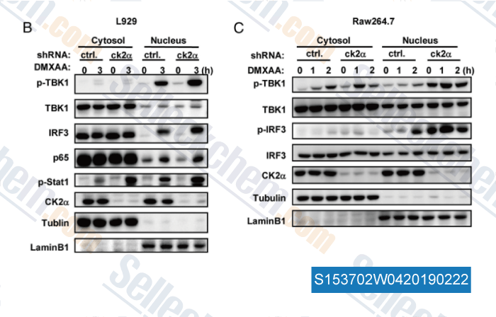
Data from [ , , J Immunol, 2015, 194:4477-4488 ]
-
Data from [ , , J Immunol, 2015, 194(9):4477-88 ]
-
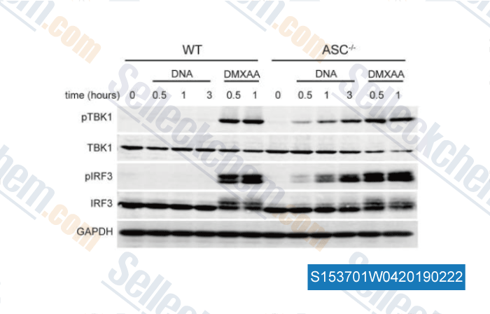
Data from [ , , J Immunol, 2016, 196(7):3191-8 ]
-
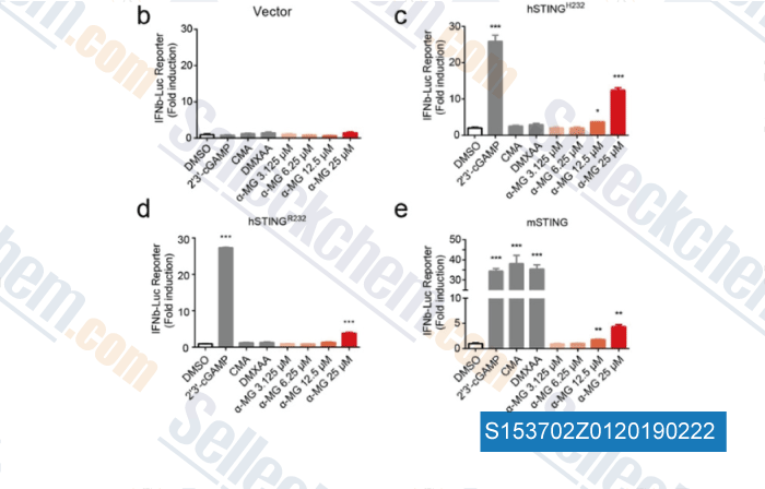
Data from [ , , ChemMedChem, 2018, 13(19):2057-2064 ]
Selleck's Vadimezan (DMXAA) has been cited by 48 publications
| An oral tricyclic STING agonist suppresses tumor growth through remodeling of the immune microenvironment [ Cell Chem Biol, 2025, 32(2):280-290.e14] | PubMed: 39904339 |
| Profile of STING agonist and inhibitor research: a bibliometric analysis [ Front Pharmacol, 2025, 16:1528459] | PubMed: 40008133 |
| C9orf72 Alleviates DSS‑Induced Ulcerative Colitis via the cGAS-STING Pathway [ Immun Inflamm Dis, 2025, 13(1):e70139] | PubMed: 39873292 |
| YTHDF2 in peritumoral hepatocytes mediates chemotherapy-induced antitumor immune responses through CX3CL1-mediated CD8+ T cell recruitment [ Mol Cancer, 2024, 23(1):186] | PubMed: 39237909 |
| S-nitrosothiol homeostasis maintained by ADH5 facilitates STING-dependent host defense against pathogens [ Nat Commun, 2024, 15(1):1750] | PubMed: 38409248 |
| Mitochondrial DNA-boosted dendritic cell-based nanovaccination triggers antitumor immunity in lung and pancreatic cancers [ Cell Rep Med, 2024, 5(7):101648] | PubMed: 38986624 |
| FMT rescues mice from DSS-induced colitis in a STING-dependent manner [ Gut Microbes, 2024, 16(1):2397879] | PubMed: 39324491 |
| TBK1-Zyxin signaling controls tumor-associated macrophage recruitment to mitigate antitumor immunity [ EMBO J, 2024, 10.1038/s44318-024-00244-9] | PubMed: 39304793 |
| ISGylation by HERCs facilitates STING activation [ Cell Rep, 2024, 43(5):114135] | PubMed: 38652662 |
| Activation of STING signaling aggravates chronic alcohol exposure-induced cognitive impairment by increasing neuroinflammation and mitochondrial apoptosis [ CNS Neurosci Ther, 2024, 30(3):e14689] | PubMed: 38516831 |
RETURN POLICY
Selleck Chemical’s Unconditional Return Policy ensures a smooth online shopping experience for our customers. If you are in any way unsatisfied with your purchase, you may return any item(s) within 7 days of receiving it. In the event of product quality issues, either protocol related or product related problems, you may return any item(s) within 365 days from the original purchase date. Please follow the instructions below when returning products.
SHIPPING AND STORAGE
Selleck products are transported at room temperature. If you receive the product at room temperature, please rest assured, the Selleck Quality Inspection Department has conducted experiments to verify that the normal temperature placement of one month will not affect the biological activity of powder products. After collecting, please store the product according to the requirements described in the datasheet. Most Selleck products are stable under the recommended conditions.
NOT FOR HUMAN, VETERINARY DIAGNOSTIC OR THERAPEUTIC USE.