|
How to Cite 1. For In-Text Citation (Materials & Methods): 2. For Key Resources Table: |
||
|
Toll Free: (877) 796-6397 -- USA and Canada only -- |
Fax: +1-832-582-8590 Orders: +1-832-582-8158 |
Tech Support: +1-832-582-8158 Ext:3 Please provide your Order Number in the email. We strive to reply to |
Technical Data
| Formula | C27H32N6 |
||||||||||
| Molecular Weight | 440.58 | CAS No. | 497839-62-0 | ||||||||
| Solubility (25°C)* | In vitro | DMSO | 88 mg/mL (199.73 mM) | ||||||||
| Ethanol | 6 mg/mL (13.61 mM) | ||||||||||
| Water | Insoluble | ||||||||||
| In vivo (Add solvents to the product individually and in order) |
|
||||||||||
|
* <1 mg/ml means slightly soluble or insoluble. * Please note that Selleck tests the solubility of all compounds in-house, and the actual solubility may differ slightly from published values. This is normal and is due to slight batch-to-batch variations. * Room temperature shipping (Stability testing shows this product can be shipped without any cooling measures.) |
|||||||||||
Preparing Stock Solutions
Biological Activity
| Description | AEE788 (NVP-AEE788) is a potent inhibitor of EGFR and HER2/ErbB2 with IC50 of 2 nM and 6 nM, respectively. It is less potent against VEGFR2/KDR, c-Abl, c-Src, and Flt-1, and does not inhibit Ins-R, IGF-1R, PKCα, or CDK1. This compound has reached Phase 1/2 clinical trials. | |||||||||||
|---|---|---|---|---|---|---|---|---|---|---|---|---|
| Targets |
|
|||||||||||
| In vitro | AEE788 (NVP-AEE788) also inhibits KDR, c-abl, c-Src, and Flt-1 with IC50 of 50-80 nM. It is not sensitive to ErbB-4, PDGFR-β, Flt-3, Flt-4, RET, and c-Kit and has no inhibitory to Ins-R, IGF-1R, PKC-α, and PKA. This compound potently inhibits EGFR phosphorylation in A431 cells with IC50 of 11 nM. It also inhibits the phosphorylation of KDR in CHO cells and erbB2 in BT-474 cells, without any effects on PDGF-induced phosphorylation in A31 cells. It inhibits the proliferation of NCI-H596, MK, BT-474 and SK-BR-3 cells with IC50 of 78, 56, 49 and 381 nM, respectively. Otherwise, it has the additional property of inhibiting cellular proliferation driven by EGFR mutant including 32D/EGFR and 32D/EGFRvIII. It further also inhibits both EGF- and VEGF-driven HUVEC proliferation with IC50 of 43 and 155 nM, respectively. It inhibits the phosphorylation of EGFR, VEGFR2, Akt, and MAPK in human cutaneous SCC cell lines (Colo16, HaCaT, SRB1, and SRB12 cells), which leads to growth inhibition and induction of apoptosis. It inhibits the phosphorylation of EGFR and Akt in HT29 cells at 0.2 to 1.0 μM. It inhibits cell proliferation and prevents EGF- and neuregulin-induced HER1, HER2, and HER3 activation in medulloblastoma cell lines. It shows growth-suppressive activities in chemosensitive and chemoresistant medulloblastoma cells. | |||||||||||
| In vivo | AEE788 (NVP-AEE788) produces a dose-dependent inhibition of tumor growth in NCI-H596 or DU145 xenograft models, with only minor body weight changes. It induces tumor regression by 57% at 50 mg/kg in the NeuT/erbB2 GeMag model and potently inhibits EGF-induced EGFR phosphorylation in A431 tumors and erbB2 phosphorylation in GeMag tumors. This compound dose-dependently inhibited angiogenesis induced by VEGF and does not inhibit bFGF-induced angiogenesis. It suppresses the growth of tumor volume by 54% in Colo16 xenografts at 50 mg/kg, which is due to the inhibition of phosphorylation of EGFR, VEGFR, Akt, and MAPK. At 50 mg/kg, it also inhibits growth of tumors in the cecum and peritoneum (>50%) and reduces the incidence of lymph node metastasis to 70% in HT29 cells implanted in the cecum of nude mice, without loss of body weight and gross evidence of neovascularization. It significantly lowers the expression levels of pEGFR and pVEGFR in HT29 cecal tumors and does not alter those of EGF, VEGF, EGFR, or VEGFR. Combined with CPT-11, it results in significantly smaller tumors and complete inhibition of lymph node metastasis. This compound inhibits the growth of Daoy, DaoyPt, and DaoyHER2 xenografts by 51%, 45%, and 72%, respectively. It could promote LBH589-mediated generation of reactive oxygen species in K562 tumor cells, which in turn increase apoptosis. |
Protocol (from reference)
| Kinase Assay:[1] |
|
|---|---|
| Cell Assay:[1] |
|
| Animal Study:[1] |
|
References
|
Customer Product Validation
-
, , The Prostate, 2013, 73:1453-1461.
-
Data from [ , , Cell Cycle, 2014, 13(15):2415-30 ]
Selleck's AEE788 (NVP-AEE788) Has Been Cited by 18 Publications
| A community challenge for a pancancer drug mechanism of action inference from perturbational profile data [ Cell Rep Med, 2022, 3(1):100492] | PubMed: 35106508 |
| Comprehensive pharmacogenomic characterization of gastric cancer. [ Genome Med, 2020, 18;12(1):17] | PubMed: 32070411 |
| Targeting the non-canonical roles of PCNA modifies and increases the response to targeted anti-cancer therapy [ Oncotarget, 2019, 10(68):7185-7197] | PubMed: 31921382 |
| Brain Distribution of a Panel of Epidermal Growth Factor Receptor Inhibitors Using Cassette Dosing in Wild-Type and Abcb1/Abcg2-Deficient Mice [ Drug Metabolism and Disposition, 2019, 393-404] | PubMed: 30705084 |
| Rationale for Using Irreversible Epidermal Growth Factor Receptor Inhibitors in Combination with Phosphatidylinositol 3-Kinase Inhibitors for Advanced Head and Neck Squamous Cell Carcinoma. [ Mol Pharmacol, 2019, 95(5):528-536] | PubMed: 30858165 |
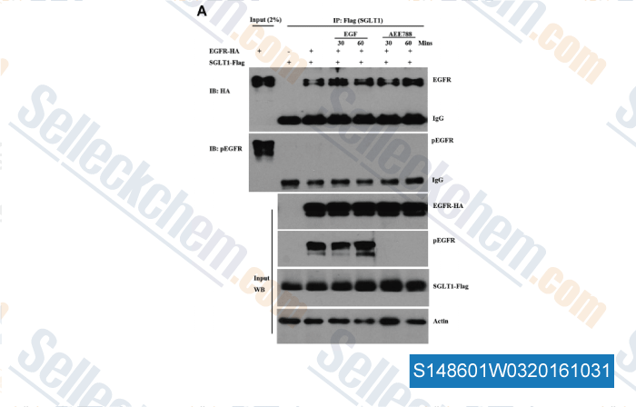
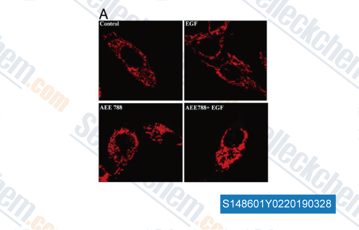
| Targeted reduction of the EGFR protein, but not inhibition of its kinase activity, induces mitophagy and death of cancer cells through activation of mTORC2 and Akt. [ Oncogenesis, 2018, 7(1):5] | PubMed: 29358623 |
| Synergistic induction of apoptosis by salinomycin and gefitinib through lysosomal and mitochondrial dependent pathway overcomes gefitinib resistance in colorectal cancer. [ Oncotarget, 2017, 8(14):22414-22432] | PubMed: 26461472 |
| A high-content EMT screen identifies multiple receptor tyrosine kinase inhibitors with activity on TGFβ receptor [ Oncotarget, 2016, 7(18-:25983-6002] | PubMed: 27036020 |
| [ Oncotarget, 2015, ] | PubMed: 25965827 |
| [ Oncotarget, 2015, ] | PubMed: 26378037 |
RETURN POLICY
Selleck Chemical’s Unconditional Return Policy ensures a smooth online shopping experience for our customers. If you are in any way unsatisfied with your purchase, you may return any item(s) within 7 days of receiving it. In the event of product quality issues, either protocol related or product related problems, you may return any item(s) within 365 days from the original purchase date. Please follow the instructions below when returning products.
SHIPPING AND STORAGE
Selleck products are transported at room temperature. If you receive the product at room temperature, please rest assured, the Selleck Quality Inspection Department has conducted experiments to verify that the normal temperature placement of one month will not affect the biological activity of powder products. After collecting, please store the product according to the requirements described in the datasheet. Most Selleck products are stable under the recommended conditions.
NOT FOR HUMAN, VETERINARY DIAGNOSTIC OR THERAPEUTIC USE.