- Bioactive Compounds
- By Signaling Pathways
- PI3K/Akt/mTOR
- Epigenetics
- Methylation
- Immunology & Inflammation
- Protein Tyrosine Kinase
- Angiogenesis
- Apoptosis
- Autophagy
- ER stress & UPR
- JAK/STAT
- MAPK
- Cytoskeletal Signaling
- Cell Cycle
- TGF-beta/Smad
- Compound Libraries
- Popular Compound Libraries
- Customize Library
- Clinical and FDA-approved Related
- Bioactive Compound Libraries
- Inhibitor Related
- Natural Product Related
- Metabolism Related
- Cell Death Related
- By Signaling Pathway
- By Disease
- Anti-infection and Antiviral Related
- Neuronal and Immunology Related
- Fragment and Covalent Related
- FDA-approved Drug Library
- FDA-approved & Passed Phase I Drug Library
- Preclinical/Clinical Compound Library
- Bioactive Compound Library-I
- Bioactive Compound Library-Ⅱ
- Kinase Inhibitor Library
- Express-Pick Library
- Natural Product Library
- Human Endogenous Metabolite Compound Library
- Alkaloid Compound LibraryNew
- Angiogenesis Related compound Library
- Anti-Aging Compound Library
- Anti-alzheimer Disease Compound Library
- Antibiotics compound Library
- Anti-cancer Compound Library
- Anti-cancer Compound Library-Ⅱ
- Anti-cancer Metabolism Compound Library
- Anti-Cardiovascular Disease Compound Library
- Anti-diabetic Compound Library
- Anti-infection Compound Library
- Antioxidant Compound Library
- Anti-parasitic Compound Library
- Antiviral Compound Library
- Apoptosis Compound Library
- Autophagy Compound Library
- Calcium Channel Blocker LibraryNew
- Cambridge Cancer Compound Library
- Carbohydrate Metabolism Compound LibraryNew
- Cell Cycle compound library
- CNS-Penetrant Compound Library
- Covalent Inhibitor Library
- Cytokine Inhibitor LibraryNew
- Cytoskeletal Signaling Pathway Compound Library
- DNA Damage/DNA Repair compound Library
- Drug-like Compound Library
- Endoplasmic Reticulum Stress Compound Library
- Epigenetics Compound Library
- Exosome Secretion Related Compound LibraryNew
- FDA-approved Anticancer Drug LibraryNew
- Ferroptosis Compound Library
- Flavonoid Compound Library
- Fragment Library
- Glutamine Metabolism Compound Library
- Glycolysis Compound Library
- GPCR Compound Library
- Gut Microbial Metabolite Library
- HIF-1 Signaling Pathway Compound Library
- Highly Selective Inhibitor Library
- Histone modification compound library
- HTS Library for Drug Discovery
- Human Hormone Related Compound LibraryNew
- Human Transcription Factor Compound LibraryNew
- Immunology/Inflammation Compound Library
- Inhibitor Library
- Ion Channel Ligand Library
- JAK/STAT compound library
- Lipid Metabolism Compound LibraryNew
- Macrocyclic Compound Library
- MAPK Inhibitor Library
- Medicine Food Homology Compound Library
- Metabolism Compound Library
- Methylation Compound Library
- Mouse Metabolite Compound LibraryNew
- Natural Organic Compound Library
- Neuronal Signaling Compound Library
- NF-κB Signaling Compound Library
- Nucleoside Analogue Library
- Obesity Compound Library
- Oxidative Stress Compound LibraryNew
- Plant Extract Library
- Phenotypic Screening Library
- PI3K/Akt Inhibitor Library
- Protease Inhibitor Library
- Protein-protein Interaction Inhibitor Library
- Pyroptosis Compound Library
- Small Molecule Immuno-Oncology Compound Library
- Mitochondria-Targeted Compound LibraryNew
- Stem Cell Differentiation Compound LibraryNew
- Stem Cell Signaling Compound Library
- Natural Phenol Compound LibraryNew
- Natural Terpenoid Compound LibraryNew
- TGF-beta/Smad compound library
- Traditional Chinese Medicine Library
- Tyrosine Kinase Inhibitor Library
- Ubiquitination Compound Library
-
Cherry Picking
You can personalize your library with chemicals from within Selleck's inventory. Build the right library for your research endeavors by choosing from compounds in all of our available libraries.
Please contact us at [email protected] to customize your library.
You could select:
- Antibodies
- Bioreagents
- qPCR
- 2x SYBR Green qPCR Master Mix
- 2x SYBR Green qPCR Master Mix(Low ROX)
- 2x SYBR Green qPCR Master Mix(High ROX)
- Protein Assay
- Protein A/G Magnetic Beads for IP
- Anti-Flag magnetic beads
- Anti-Flag Affinity Gel
- Anti-Myc magnetic beads
- Anti-HA magnetic beads
- Poly FLAG Peptide lyophilized powder
- Protease Inhibitor Cocktail
- Protease Inhibitor Cocktail (EDTA-Free, 100X in DMSO)
- Phosphatase Inhibitor Cocktail (2 Tubes, 100X)
- Cell Biology
- Cell Counting Kit-8 (CCK-8)
- Animal Experiment
- Mouse Direct PCR Kit (For Genotyping)
- New Products
- Contact Us
Prostaglandin E2 (PGE2)
Synonyms: Dinoprostone
Prostaglandin E2 (PGE2, Dinoprostone) plays important effects in labour (softens cervix and causes uterine contraction) and also stimulates osteoblasts to release factors that stimulate bone resorption by osteoclasts.

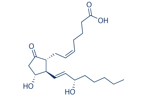
Prostaglandin E2 (PGE2) Chemical Structure
CAS: 363-24-6
Selleck's Prostaglandin E2 (PGE2) has been cited by 17 publications
Purity & Quality Control
Batch:
Purity: >97%
97
Prostaglandin E2 (PGE2) Related Products
| Related Compound Libraries | FDA-approved Drug Library Natural Product Library Bioactive Compound Library-I Exosome Secretion Related Compound Library Human Hormone Related Compound Library | Click to Expand |
|---|
Choose Selective PGES Inhibitors
Cell Data
| Cell Lines | Assay Type | Concentration | Incubation Time | Formulation | Activity Description | PMID |
|---|---|---|---|---|---|---|
| HeLa | Function assay | TP_TRANSPORTER: uptake in PGT-expressing HeLa cells, Km = 0.094 μM. | 7754369 | |||
| HeLa | Function assay | TP_TRANSPORTER: uptake in PGT-expressing HeLa cells, K1/2 = 0.1 μM. | 8787677 | |||
| S2 | Function assay | TP_TRANSPORTER: uptake in OCT2-expressing S2 cells, Km = 0.0289 μM. | 11907186 | |||
| S2 | Function assay | TP_TRANSPORTER: uptake in OAT4-expressing S2 cells, Km = 0.154 μM. | 11907186 | |||
| S2 | Function assay | TP_TRANSPORTER: uptake in OAT3-expressing S2 cells, Km = 0.345 μM. | 11907186 | |||
| S2 | Function assay | TP_TRANSPORTER: uptake in OCT1-expressing S2 cells, Km = 0.657 μM. | 11907186 | |||
| S2 | Function assay | TP_TRANSPORTER: uptake in OAT2-expressing S2 cells, Km = 0.713 μM. | 11907186 | |||
| S2 | Function assay | TP_TRANSPORTER: uptake in OAT1-expressing S2 cells, Km = 0.97 μM. | 11907186 | |||
| HEK293 | Function assay | Inhibitory activity against human EP4 receptor expressed in HEK293 ebna cells, IC50 = 0.0007 μM. | 12643927 | |||
| HEK293 | Function assay | EP4 agonist potency utilizing a stable clone of pSV40-EP4 transfected into HEK293 cells expressing EP4 receptor, EC50 = 0.003 μM. | 12643927 | |||
| Sf9 | Function assay | TP_TRANSPORTER: uptake (vesicle) in membrane vesicles from MRP4-expressing Sf9 cells, Km = 3.4 μM. | 12835412 | |||
| HEK293 | Function assay | Displacement of [3H]PGE2 from human EP3 receptor expressed in HEK293 cells, Ki = 0.00033 μM. | 17531488 | |||
| HEK293 | Function assay | Displacement of [3H]PGE2 from human EP4 receptor expressed in HEK293 cells, Ki = 0.00079 μM. | 17531488 | |||
| HEK293 | Function assay | Displacement of [3H]PGE2 from human EP2 receptor expressed in HEK293 cells, Ki = 0.0049 μM. | 17531488 | |||
| HEK293 | Function assay | Displacement of [3H]PGE2 from human EP1 receptor expressed in HEK293 cells, Ki = 0.0091 μM. | 17531488 | |||
| HEK293 | Function assay | Agonist activity against rat EP2 receptor expressed in HEK293 cells assessed as stimulation of cAMP release, EC50 = 0.0002 μM. | 19250823 | |||
| HEK293 | Function assay | Agonist activity against rat EP4 receptor expressed in HEK293 cells assessed as stimulation of cAMP release, EC50 = 0.0007 μM. | 19250823 | |||
| HEK293 | Function assay | Inhibition of rat EP4 receptor expressed in HEK293 cells, IC50 = 0.0021 μM. | 19250823 | |||
| HEK293 | Function assay | Inhibition of rat EP2 receptor expressed in HEK293 cells, IC50 = 0.0052 μM. | 19250823 | |||
| CHO | Function assay | 60 mins | Displacement of [3H]-PGE2 from mouse EP4 receptor expressed in CHO cells after 60 mins by liquid scintillation counting, Ki = 0.0031 μM. | 22119471 | ||
| CHO | Function assay | 60 mins | Displacement of [3H]-PGE2 from mouse EP3 receptor expressed in CHO cells after 60 mins by liquid scintillation counting, Ki = 0.005 μM. | 22119471 | ||
| CHO | Function assay | 20 mins | Displacement of [3H]-PGE2 from mouse EP1 receptor expressed in CHO cells after 20 mins by liquid scintillation counting, Ki = 0.006 μM. | 22119471 | ||
| CHO | Function assay | 60 mins | Displacement of [3H]-PGE2 from mouse EP2 receptor expressed in CHO cells after 60 mins by liquid scintillation counting, Ki = 0.022 μM. | 22119471 | ||
| CHO | Function assay | 60 mins | Displacement of [3H]-PGE2 from mouse EP4 receptor expressed in CHO cells after 60 mins by scintillation counting, Ki = 0.0031 μM. | 22386979 | ||
| CHO | Function assay | 60 mins | Displacement of [3H]-PGE2 from mouse EP3 receptor expressed in CHO cells after 60 mins by scintillation counting, Ki = 0.005 μM. | 22386979 | ||
| CHO | Function assay | 60 mins | Displacement of [3H]-PGE2 from mouse EP1 receptor expressed in CHO cells after 60 mins by scintillation counting, Ki = 0.006 μM. | 22386979 | ||
| CHO | Function assay | 60 mins | Displacement of [3H]-PGE2 from mouse EP2 receptor expressed in CHO cells after 60 mins by scintillation counting, Ki = 0.022 μM. | 22386979 | ||
| CHEM1 | Function assay | 2 hrs | Displacement of [3H]PGE2 from human recombinant prostanoid EP4 receptor in CHEM1 cells after 2 hrs, Ki = 0.00045 μM. | 23403082 | ||
| CHEM1 | Function assay | 2 hrs | Displacement of [3H]PGE2 from human recombinant prostanoid EP4 receptor in CHEM1 cells after 2 hrs, IC50 = 0.0011 μM. | 23403082 | ||
| CHO | Function assay | 100 nM | Activity at recombinant EP1 receptor (unknown origin) expressed in CHO cells co-expressing Gq protein at 100 nM by electrical cell substrate impedance sensing system | 25701254 | ||
| CHO | Function assay | 100 nM | Activity at recombinant EP4 receptor (unknown origin) expressed in CHO cells co-expressing Gs protein at 100 nM by electrical cell substrate impedance sensing system | 25701254 | ||
| CHO | Function assay | 100 nM | Activity at recombinant EP3 receptor (unknown origin) expressed in CHO cells co-expressing Gi protein at 100 nM by electrical cell substrate impedance sensing system | 25701254 | ||
| CHO | Function assay | 30 mins | Agonist activity at human EP2 receptor expressed in CHO cells assessed as increase in intracellular cAMP level after 30 mins by HTRF method, EC50 = 0.0019 μM. | 26985320 | ||
| CHO | Function assay | Agonist activity at human EP3 receptor expressed in CHO cells assessed as increase in intracellular calcium level by fluorescence based analysis, EC50 = 0.0025 μM. | 26985320 | |||
| CHO | Function assay | Agonist activity at human EP1 receptor expressed in CHO cells assessed as increase in intracellular calcium level by fluorescence based analysis, EC50 = 0.0037 μM. | 26985320 | |||
| CHO | Function assay | 30 mins | Agonist activity at human EP4 receptor expressed in CHO cells assessed as increase in intracellular cAMP level after 30 mins by HTRF method, EC50 = 0.0075 μM. | 26985320 | ||
| Chem1 | Function assay | Agonist activity at human FP receptor expressed in human Chem1 cells assessed as increase in intracellular calcium level by fluorescence based analysis, EC50 = 0.25 μM. | 26985320 | |||
| HEK293 | Function assay | 90 mins | Agonist activity at PK2-tagged human EP2 receptor expressed in HEK293 cells assessed as induction of EA-tagged beta-arrestin recruitment incubated for 90 mins by beta-galactosidase reporter gene assay, EC50 = 0.346 μM. | 26985320 | ||
| CHO | Function assay | 30 mins | Agonist activity at human IP receptor expressed in CHO cells assessed as increase in intracellular cAMP level after 30 mins by HTRF method, EC50 = 0.347 μM. | 26985320 | ||
| HEK293 | Function assay | Displacement of [3H]PGE2 from human recombinant prostanoid EP2 receptor expressed in HEK293 cells, IC50 = 0.0026 μM. | 26988801 | |||
| HEK293 | Function assay | 120 mins | Displacement of [3H]PGE2 from human recombinant EP4 receptor expressed in HEK293 cells measured after 120 mins by scintillation counting method, IC50 = 0.00055 μM. | 27876250 | ||
| HEK293 | Function assay | 120 mins | Displacement of [3H]PGE2 from human recombinant EP2 receptor expressed in HEK293 cells measured after 120 mins by scintillation counting method, IC50 = 0.0026 μM. | 27876250 | ||
| Click to View More Cell Line Experimental Data | ||||||
Biological Activity
| Description | Prostaglandin E2 (PGE2, Dinoprostone) plays important effects in labour (softens cervix and causes uterine contraction) and also stimulates osteoblasts to release factors that stimulate bone resorption by osteoclasts. |
|---|
| In vitro | ||||
| In vitro | PGE2 acts via EP4 receptors on osteoclasts during the progression of OA and OA-related pain.[1] |
|||
|---|---|---|---|---|
| Cell Research | Cell lines | Mice osteoclasts differentiated from bone marrow macrophages (BMMs) | ||
| Concentrations | 1, 10, 100, 1000, 10000 nM | |||
| Incubation Time | 18 h | |||
| Method | When BMMs differentiated into mature osteoclasts, a TRAP staining kit is used to stain mature osteoclasts according to the manufacturer s instructions, and TRAP-positive osteoclasts with 5 or more nuclei are counted. For BMM migration, BMMs (1×10⁴ cells per well) are seeded in the upper chamber of the Transwell inserts with or without inhibitors and antagonists. PGE2 is added in lower chamber. The culture medium is the same between the upper chamber and lower chamber in the Transwell. After 18 h, the cells are fixed with 4% PFA and stained with crystal violet. The cells on the bottom of the Transwell insert are used to assess migration. At least five fields of view per insert are photographed, and migrated cells are counted. |
|||
| In Vivo | ||
| In vivo |
PGE2 (0.3 μg/k, i.p.) significantly reduces the number of peritoneab macrophages undergoing phagocytosis of the methacrybate microbeads in rats. |
|
|---|---|---|
| Animal Research | Animal Models | Male Sprague Dawley rats |
| Dosages | 0.3 µg/kg | |
| Administration | i.p. | |
| NCT Number | Recruitment | Conditions | Sponsor/Collaborators | Start Date | Phases |
|---|---|---|---|---|---|
| NCT06129604 | Not yet recruiting | Colorectal Carcinoma (CRC)|Endometrial Carcinoma (EC) |
University of Oklahoma |
January 2024 | Phase 2 |
| NCT06190132 | Completed | Uremic Pruritus in Hemodialysis Patients |
Ain Shams University |
April 1 2023 | Not Applicable |
| NCT05412810 | Recruiting | Mechanical Ventilation Complication|Hyperoxia |
Ohio State University |
March 1 2023 | -- |
Chemical Information & Solubility
| Molecular Weight | 352.47 | Formula | C20H32O5 |
| CAS No. | 363-24-6 | SDF | Download Prostaglandin E2 (PGE2) SDF |
| Smiles | CCCCCC(C=CC1C(CC(=O)C1CC=CCCCC(=O)O)O)O | ||
| Storage (From the date of receipt) | |||
|
In vitro |
DMSO : 70 mg/mL ( (198.59 mM); Moisture-absorbing DMSO reduces solubility. Please use fresh DMSO.) Ethanol : 70 mg/mL Water : 2.5 mg/mL |
Molecular Weight Calculator |
|
In vivo Add solvents to the product individually and in order. |
In vivo Formulation Calculator |
||||
Preparing Stock Solutions
Molarity Calculator
In vivo Formulation Calculator (Clear solution)
Step 1: Enter information below (Recommended: An additional animal making an allowance for loss during the experiment)
mg/kg
g
μL
Step 2: Enter the in vivo formulation (This is only the calculator, not formulation. Please contact us first if there is no in vivo formulation at the solubility Section.)
% DMSO
%
% Tween 80
% ddH2O
%DMSO
%
Calculation results:
Working concentration: mg/ml;
Method for preparing DMSO master liquid: mg drug pre-dissolved in μL DMSO ( Master liquid concentration mg/mL, Please contact us first if the concentration exceeds the DMSO solubility of the batch of drug. )
Method for preparing in vivo formulation: Take μL DMSO master liquid, next addμL PEG300, mix and clarify, next addμL Tween 80, mix and clarify, next add μL ddH2O, mix and clarify.
Method for preparing in vivo formulation: Take μL DMSO master liquid, next add μL Corn oil, mix and clarify.
Note: 1. Please make sure the liquid is clear before adding the next solvent.
2. Be sure to add the solvent(s) in order. You must ensure that the solution obtained, in the previous addition, is a clear solution before proceeding to add the next solvent. Physical methods such
as vortex, ultrasound or hot water bath can be used to aid dissolving.
Tech Support
Answers to questions you may have can be found in the inhibitor handling instructions. Topics include how to prepare stock solutions, how to store inhibitors, and issues that need special attention for cell-based assays and animal experiments.
Tel: +1-832-582-8158 Ext:3
If you have any other enquiries, please leave a message.
* Indicates a Required Field
Tags: buy Prostaglandin E2 (PGE2) | Prostaglandin E2 (PGE2) ic50 | Prostaglandin E2 (PGE2) price | Prostaglandin E2 (PGE2) cost | Prostaglandin E2 (PGE2) solubility dmso | Prostaglandin E2 (PGE2) purchase | Prostaglandin E2 (PGE2) manufacturer | Prostaglandin E2 (PGE2) research buy | Prostaglandin E2 (PGE2) order | Prostaglandin E2 (PGE2) mouse | Prostaglandin E2 (PGE2) chemical structure | Prostaglandin E2 (PGE2) mw | Prostaglandin E2 (PGE2) molecular weight | Prostaglandin E2 (PGE2) datasheet | Prostaglandin E2 (PGE2) supplier | Prostaglandin E2 (PGE2) in vitro | Prostaglandin E2 (PGE2) cell line | Prostaglandin E2 (PGE2) concentration | Prostaglandin E2 (PGE2) nmr


