
- Bioactive Compounds
- By Signaling Pathways
- PI3K/Akt/mTOR
- Epigenetics
- Methylation
- Immunology & Inflammation
- Protein Tyrosine Kinase
- Angiogenesis
- Apoptosis
- Autophagy
- ER stress & UPR
- JAK/STAT
- MAPK
- Cytoskeletal Signaling
- Cell Cycle
- TGF-beta/Smad
- Compound Libraries
- Antibodies
- Bioreagents
- qPCR
- 2x SYBR Green qPCR Master Mix
- 2x SYBR Green qPCR Master Mix(Low ROX)
- 2x SYBR Green qPCR Master Mix(High ROX)
- Protein Assay
- Protein A/G Magnetic Beads for IP
- Anti-Flag magnetic beads
- Anti-Flag Affinity Gel
- Anti-Myc magnetic beads
- Anti-HA magnetic beads
- Poly FLAG Peptide lyophilized powder
- Protease Inhibitor Cocktail
- Protease Inhibitor Cocktail (EDTA-Free, 100X in DMSO)
- Phosphatase Inhibitor Cocktail (2 Tubes, 100X)
- Cell Biology
- Cell Counting Kit-8 (CCK-8)
- Animal Experiment
- Mouse Direct PCR Kit (For Genotyping)
- New Products
- Contact Us
-
 Australia
-
 Austria
-
 Belgium
-
 Brazil
-
 Canada
-
 China
China
-
 Czech Republic
-
 Denmark
-
 Finland
-
 France
-
 Germany
-
 Greece
-
 Hong Kong
-
 Hungary
-
 Iceland
-
 India
India
-
 Ireland
-
 Israel
-
 Italy
Italy
-
 Japan
Japan
-
 Korea
Korea
-
 Luxembourg
-
 Malaysia
-
 Netherlands
-
 New Zealand
-
 Norway
-
 Poland
-
 Qatar
Qatar
-
 Romania
-
 Saudi Arabia
-
 Singapore
-
 Spain
Spain
-
 Sweden
-
 Switzerland
-
 Taiwan
-
 Turkey
-
 United Kingdom
-
 United States
-
 Other Countries
3PO
Synonyms: 3-(3-pyridinyl)-1-(4-pyridinyl)-2-propen-1-one
3PO (3-(3-pyridinyl)-1-(4-pyridinyl)-2-propen-1-one) is a small-molecule inhibitor of PFKFB3 with an IC50 of 22.9 μM for recombinant human PFKFB3 protein and does not inhibit PFK-1 activity. It suppresses glucose uptake, and decreases the intracellular concentration of Fru-2,6-BP, lactate, ATP, NAD+, and NADH.
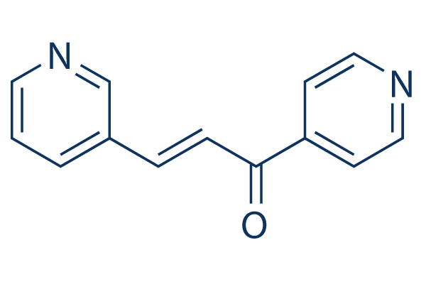
3PO Chemical Structure
CAS: 18550-98-6
Selleck's 3PO has been cited by 7 publications
Purity & Quality Control
Batch:
Purity:
99.94%
99.94
3PO Related Products
| Related Targets | PFKFB3 PFKFB4 | Click to Expand |
|---|---|---|
| Related Compound Libraries | Kinase Inhibitor Library PI3K/Akt Inhibitor Library MAPK Inhibitor Library DNA Damage/DNA Repair compound Library Cell Cycle compound library | Click to Expand |
Choose Selective PFKFB Inhibitors
Cell Data
| Cell Lines | Assay Type | Concentration | Incubation Time | Formulation | Activity Description | PMID |
|---|---|---|---|---|---|---|
| Jurkat | Growth inhibition assay | 48 hrs | Growth inhibition of human Jurkat cells expressing inducible FLAG-tagged PFKFB incubated for 48 hrs by trypan blue dye exclusion assay, IC50=1.4μM | ChEMBL | ||
| NHBE | Growth inhibition assay | 48 to 72 hrs | Growth inhibition of human NHBE cells immortalized with human telomerase and large-T antigen and transformed with mutated Ras protein incubated for 48 to 72 hrs by trypan blue dye exclusion assay, IC50=1.5μM | ChEMBL | ||
| K562 | Growth inhibition assay | 48 hrs | Growth inhibition of human K562 cells incubated for 48 hrs by trypan blue dye exclusion assay, IC50=3.2μM | ChEMBL | ||
| HL60 | Growth inhibition assay | 48 hrs | Growth inhibition of human HL60 cells incubated for 48 hrs by trypan blue dye exclusion assay, IC50=4.5μM | ChEMBL | ||
| MDA-MB-231 | Growth inhibition assay | 48 hrs | Growth inhibition of human MDA-MB-231 cells incubated for 48 hrs by trypan blue dye exclusion assay, IC50=4.7μM | ChEMBL | ||
| Jurkat | Growth inhibition assay | 48 hrs | Growth inhibition of human Jurkat cells expressing un-altered PFKFB expression incubated for 48 hrs by trypan blue dye exclusion assay, IC50=8.9μM | ChEMBL | ||
| CRL11174 | Growth inhibition assay | 48 hrs | Growth inhibition of human CRL11174 cells incubated for 48 hrs by trypan blue dye exclusion assay, IC50=15μM | ChEMBL | ||
| LLC | Growth inhibition assay | 48 hrs | Growth inhibition of mouse LLC cells incubated for 48 hrs by trypan blue dye exclusion assay, IC50=19μM | ChEMBL | ||
| Jurkat | Growth inhibition assay | 48 hrs | Growth inhibition of human Jurkat cells expressing inducible FLAG-tagged PFKFB in presence of doxycycline incubated for 48 hrs by trypan blue dye exclusion assay, IC50=19.3μM | ChEMBL | ||
| HeLa | Growth inhibition assay | 48 hrs | Growth inhibition of human HeLa cells incubated for 48 hrs by trypan blue dye exclusion assay, IC50=24μM | ChEMBL | ||
| Jurkat | Growth inhibition assay | 48 hrs | Growth inhibition of PFKFB3+/-ht/LT/Ras human Jurkat cells and incubated for 48 hrs by trypan blue dye exclusion assay, IC50=26μM | ChEMBL | ||
| A549 | Growth inhibition assay | 48 hrs | Growth inhibition of human A549 cells incubated for 48 hrs by trypan blue dye exclusion assay, IC50=48μM | ChEMBL | ||
| Jurkat | Growth inhibition assay | 48 hrs | Growth inhibition of PFKFB3+/+ht/LT/Ras human Jurkat cells and incubated for 48 hrs by trypan blue dye exclusion assay, IC50=49μM | ChEMBL | ||
| Jurkat | Cell proliferation assay | 10 uM | 36 hrs | Inhibition of cell proliferation of human Jurkat cells expressing inducible FLAG-tagged PFKFB at 10 uM incubated for 36 hrs by trypan blue dye exclusion assay | ChEMBL | |
| Jurkat | Cell cycle assay | Inhibition of cell cycle arrest in human Jurkat cells expressing inducible FLAG-tagged PFKFB assessed as accumulation at G2/M phase by propidium iodide staining based flow cytometry | ChEMBL | |||
| Jurkat | Function assay | 10 uM | Reduction in 2-deoxy-glucose uptake in human Jurkat cells expressing inducible FLAG-tagged PFKFB at 10 uM measured within 4 hrs using [14C]-2-deoxy-glucose by scintillation counting method | ChEMBL | ||
| Jurkat | Function assay | 10 uM | 4 hrs | Reduction in Fru-2,6-BP production in human Jurkat cells expressing inducible FLAG-tagged PFKFB at 10 uM measured within 4 hrs | ChEMBL | |
| Jurkat | Function assay | 10 uM | 8 hrs | Reduction in lactate secretion in human Jurkat cells expressing inducible FLAG-tagged PFKFB at 10 uM after 8 hrs by lactate oxidase based colorimetric assay | ChEMBL | |
| Jurkat | Function assay | 10 uM | 16 hrs | Reduction in NADH level in human Jurkat cells expressing inducible FLAG-tagged PFKFB at 10 uM after 16 hrs | ChEMBL | |
| Jurkat | Function assay | 10 uM | 24 hrs | Reduction in NAD+ level in human Jurkat cells expressing inducible FLAG-tagged PFKFB at 10 uM after 24 hrs | ChEMBL | |
| Jurkat | Function assay | 10 uM | 24 hrs | Reduction in ATP level in human Jurkat cells expressing inducible FLAG-tagged PFKFB at 10 uM after 24 hrs | ChEMBL | |
| Jurkat | Function assay | 10 uM | 36 hrs | Suppression of glycolytic flux into lactate in human Jurkat cells expressing inducible FLAG-tagged PFKFB at 10 uM after 36 hrs by NMR spectroscopy | ChEMBL | |
| NHBE | Growth inhibition assay | 1 uM | 48 to 72 hrs | Growth inhibition of human NHBE cells immortalized with human telomerase and large-T antigen and transformed with mutated Ras protein at 1 uM incubated for 48 to 72 hrs by trypan blue dye exclusion assay | ChEMBL | |
| NHBE | Growth inhibition assay | 10 uM | 48 to 72 hrs | Growth inhibition of human NHBE cells immortalized with human telomerase and large-T antigen and transformed with mutated Ras protein at 10 uM incubated for 48 to 72 hrs by trypan blue dye exclusion assay | ChEMBL | |
| Click to View More Cell Line Experimental Data | ||||||
Biological Activity
| Description | 3PO (3-(3-pyridinyl)-1-(4-pyridinyl)-2-propen-1-one) is a small-molecule inhibitor of PFKFB3 with an IC50 of 22.9 μM for recombinant human PFKFB3 protein and does not inhibit PFK-1 activity. It suppresses glucose uptake, and decreases the intracellular concentration of Fru-2,6-BP, lactate, ATP, NAD+, and NADH. | ||
|---|---|---|---|
| Targets |
|
| In vitro | ||||
| In vitro | 3PO is an inhibitor of the PFKFB3 isozyme primarily through competition with Fru-6-P and does not inhibit purified PFK-1 activity. 3PO markedly attenuates the proliferation of several human malignant hematopoietic and adenocarcinoma cell lines (IC50, 1.4-24 μmol/L) and is selectively cytostatic to ras-transformed human bronchial epithelial cells relative to normal human bronchial epithelial cells. 3PO can cause G2-M phase arrest[1]. | |||
|---|---|---|---|---|
| Cell Research | Cell lines | Jurkat cells | ||
| Concentrations | 10 μmol/L | |||
| Incubation Time | 0, 4, 8, 16, 24, or 36 h | |||
| Method | Jurkat cells are plated at 1 × 10⁵/mL in RPMI 1640 supplemented with 10% fetal bovine serum and 50 μg/mL gentamicin sulfate. Cells are immediately treated with vehicle or 10 μmol/L 3PO for 0, 4, 8, 16, 24, or 36 h. Cell cycle analysis is done. |
|||
| In Vivo | ||
| In vivo | i.p. administration of 3PO (0.07 mg/g) to tumor-bearing mice markedly reduces the intracellular concentration of Fru-2,6-BP, glucose uptake, and growth of established tumors in vivo. It suppresses tumorigenic growth of breast adenocarcinoma, leukemia, and lung adenocarcinoma cells in vivo[1]. The PK properties of 3PO are examined in C57Bl/6 mice intravenously administered 3PO: clearance CL=2312 mL/min/kg, T1/2=0.3 hr, Cmax=113 ng/ml, AUC0-inf=36 ng/hr/ml. 3PO is reported to have potent activity against a highly relevant mouse model of leukemia[2]. | |
|---|---|---|
| Animal Research | Animal Models | tumor bearing mice (BALB/c nude mice or C57Bl/6 female mice background) |
| Dosages | 0.07 mg/g | |
| Administration | i.p. | |
Chemical Information & Solubility
| Molecular Weight | 210.23 | Formula | C13H10N2O |
| CAS No. | 18550-98-6 | SDF | Download 3PO SDF |
| Smiles | C1=CC(=CN=C1)C=CC(=O)C2=CC=NC=C2 | ||
| Storage (From the date of receipt) | |||
|
In vitro |
DMSO : 42 mg/mL ( (199.78 mM); Moisture-absorbing DMSO reduces solubility. Please use fresh DMSO.) Ethanol : 11 mg/mL Water : Insoluble |
Molecular Weight Calculator |
|
In vivo Add solvents to the product individually and in order. |
In vivo Formulation Calculator |
||||
Preparing Stock Solutions
Molarity Calculator
In vivo Formulation Calculator (Clear solution)
Step 1: Enter information below (Recommended: An additional animal making an allowance for loss during the experiment)
mg/kg
g
μL
Step 2: Enter the in vivo formulation (This is only the calculator, not formulation. Please contact us first if there is no in vivo formulation at the solubility Section.)
% DMSO
%
% Tween 80
% ddH2O
%DMSO
%
Calculation results:
Working concentration: mg/ml;
Method for preparing DMSO master liquid: mg drug pre-dissolved in μL DMSO ( Master liquid concentration mg/mL, Please contact us first if the concentration exceeds the DMSO solubility of the batch of drug. )
Method for preparing in vivo formulation: Take μL DMSO master liquid, next addμL PEG300, mix and clarify, next addμL Tween 80, mix and clarify, next add μL ddH2O, mix and clarify.
Method for preparing in vivo formulation: Take μL DMSO master liquid, next add μL Corn oil, mix and clarify.
Note: 1. Please make sure the liquid is clear before adding the next solvent.
2. Be sure to add the solvent(s) in order. You must ensure that the solution obtained, in the previous addition, is a clear solution before proceeding to add the next solvent. Physical methods such
as vortex, ultrasound or hot water bath can be used to aid dissolving.
Tech Support
Answers to questions you may have can be found in the inhibitor handling instructions. Topics include how to prepare stock solutions, how to store inhibitors, and issues that need special attention for cell-based assays and animal experiments.
Tel: +1-832-582-8158 Ext:3
If you have any other enquiries, please leave a message.
* Indicates a Required Field
Tags: buy 3PO | 3PO supplier | purchase 3PO | 3PO cost | 3PO manufacturer | order 3PO | 3PO distributor
Products are for research use only. Not for human use. We do not sell to patients.
©Copyright 2013 Selleck Chemicals. All Rights Reserved.